

A stable Lisfranc sprain can heal without surgery — the right boot and progression schedule is the whole game.
You are in the right place. Dr. Tom Biernacki, DPM, FACFAS — board-certified foot & ankle surgeon with 3,000+ surgeries — explains exactly what Lisfranc injury — conservative treatment for stable midfoot sprain means and what works. Call (810) 206-1402 for same-day appointment at Howell or Bloomfield Township.
Quick answer: Treatment for lisfranc injury conservative treatment stable midfoot sprain follows a stepwise approach: 1) conservative care first (rest, ice, supportive footwear, OTC anti-inflammatories), 2) physical therapy and targeted exercises, 3) in-office treatments (injections, custom orthotics) if conservative fails at 4-6 weeks, 4) surgery for refractory cases. Most patients resolve at step 1 or 2. Call (810) 206-1402.
Medically reviewed by Dr. Tom Biernacki, DPM — Board-Certified Podiatric Surgeon — Balance Foot & Ankle, Howell & Bloomfield Township, MI. Last updated April 2026.
Medically Reviewed by Dr. Tom Biernacki, DPM — Board-Certified Podiatrist, Balance Foot & Ankle Specialists, Michigan. Last updated April 2026.
Medically Reviewed by Dr. Tom Biernacki, DPM · Board-Certified Podiatrist · Balance Foot & Ankle Specialists · Last updated April 2026
⚡ Quick Answer
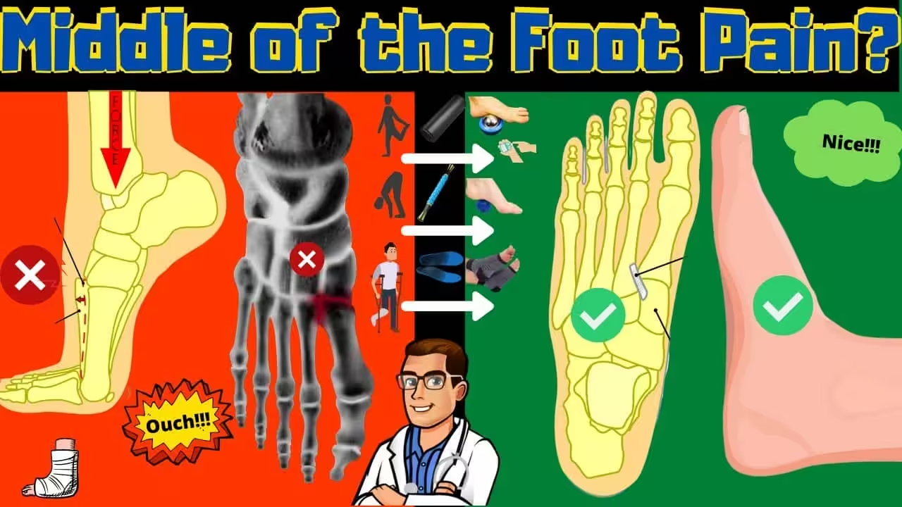
A Lisfranc injury (tarsometatarsal joint injury) can be treated without surgery when the injury is stable — meaning weight-bearing X-rays show less than 2mm of displacement and no diastasis between the first and second metatarsal bases. Conservative treatment involves 6-8 weeks of strict non-weight-bearing in a cast or boot followed by 4-6 weeks of progressive weight-bearing with structured orthotic support. The critical requirement is confirming stability through weight-bearing radiographs, as unstable Lisfranc injuries treated conservatively have unacceptable rates of midfoot collapse, chronic pain, and post-traumatic arthritis. Approximately 40-50% of Lisfranc injuries are stable enough for non-surgical management.
Table of Contents
- What Is a Lisfranc Injury?
- Lisfranc Joint Complex Anatomy
- Stable vs. Unstable: Determining Treatment Path
- Conservative Treatment Protocol
- Weight-Bearing Progression Timeline
- Rehabilitation and Return to Activity
- When Surgery Becomes Necessary
- Long-Term Outcomes of Conservative Treatment
- Complete Lisfranc Recovery Kit
- Most Common Mistake
- Warning Signs
- Watch: Lisfranc Injury Explained
- Frequently Asked Questions
- Sources
- Schedule Your Evaluation
Affiliate disclosure: This article contains affiliate links to products we recommend. As an Amazon Associate, we earn from qualifying purchases at no additional cost to you. We only recommend products we use in clinical practice and believe will benefit our patients.
What Is a Lisfranc Injury? Understanding Midfoot Joint Disruption
A Lisfranc injury involves damage to the ligaments and/or bones at the tarsometatarsal (TMT) joint complex — the articulation between the midfoot tarsal bones (cuneiforms and cuboid) and the bases of the five metatarsal bones. Named after Jacques Lisfranc de Saint-Martin, a French surgeon in Napoleon’s army who described amputations through this joint, the Lisfranc joint complex is the critical transition point between the rigid rearfoot and the flexible forefoot. Its structural integrity is essential for normal push-off during walking and running.
Lisfranc injuries exist on a spectrum from mild ligament sprains to complete fracture-dislocations. The injury is notoriously underdiagnosed — studies suggest that up to 20% of Lisfranc injuries are missed on initial evaluation, often dismissed as simple midfoot sprains. This diagnostic delay is particularly problematic because even mild-appearing Lisfranc injuries can produce devastating long-term consequences (chronic pain, midfoot collapse, post-traumatic arthritis) if instability is present and not appropriately treated.
In our Southeast Michigan practice, we see Lisfranc injuries from diverse mechanisms: athletes who sustain axial loading injuries during football or soccer, weekend warriors who catch their foot in a hole while running, and everyday patients who miss a step or twist their foot on uneven ground. The winter months bring an increase in Lisfranc injuries from slipping on ice — the foot catches while the body’s momentum continues forward, creating the classic indirect plantarflexion-axial loading mechanism that disrupts the Lisfranc ligament complex.
Lisfranc Joint Complex Anatomy: The Keystone Architecture
The Lisfranc joint complex is an architectural marvel that combines bony interlocking (like a Roman arch keystone) with powerful ligamentous support to create a structure that is simultaneously rigid enough for push-off and flexible enough for terrain adaptation. Understanding this anatomy is essential for appreciating why even small disruptions can produce significant functional impairment.
The keystone of the Lisfranc complex is the second metatarsal base, which is recessed approximately 8mm between the medial and lateral cuneiforms, creating a mortise configuration that provides inherent bony stability. This recessed position makes the second TMT joint the most stable in the complex and explains why the second metatarsal base is the last structure to displace in progressive Lisfranc disruptions — and often the first place where subtle instability is detected on imaging.
The Lisfranc ligament proper is a strong plantar ligament that connects the medial cuneiform to the second metatarsal base. It is the primary stabilizer of the medial column of the midfoot and the most important ligamentous structure in the complex. There is no corresponding dorsal ligament of equivalent strength — the dorsal ligaments are weaker than the plantar ligaments throughout the TMT complex — which is why Lisfranc dislocations characteristically displace dorsally (upward) rather than plantarly. Disruption of the Lisfranc ligament creates the hallmark diastasis (widening) between the first and second metatarsal bases that is the radiographic signature of instability.
Stable vs. Unstable: Determining Your Treatment Path
The single most critical decision in Lisfranc injury management is determining whether the injury is stable or unstable. This distinction directly determines whether conservative treatment will succeed or whether surgical fixation is required. Getting this determination wrong has serious consequences — an unstable Lisfranc injury treated conservatively will inevitably develop midfoot collapse, chronic pain, and accelerated arthritis that requires salvage surgery (arthrodesis) under far less favorable conditions than primary fixation.
Weight-bearing radiographs are the essential diagnostic study for determining stability. Non-weight-bearing X-rays can appear deceptively normal because the absence of body weight allows the disrupted bones to fall back into near-anatomic alignment. When the patient bears weight on the injured foot, unstable injuries reveal themselves through displacement — even 1-2mm of widening between the first and second metatarsal bases on the AP weight-bearing view, or dorsal displacement of metatarsal bases on the lateral weight-bearing view, indicates instability that is unlikely to respond to conservative management.
Stability criteria for conservative treatment: A Lisfranc injury is considered stable and appropriate for conservative management when weight-bearing radiographs demonstrate less than 2mm of diastasis between the first and second metatarsal bases, no dorsal subluxation on the lateral view, alignment of the medial border of the second metatarsal with the medial border of the middle cuneiform (Lisfranc alignment line intact), and no fractures involving the articular surfaces. If any of these criteria are not met, surgical fixation should be strongly considered regardless of how “minor” the injury appears clinically.
MRI provides additional information about ligament integrity when weight-bearing radiographs are equivocal. MRI can directly visualize the Lisfranc ligament and assess whether it is intact, partially torn, or completely disrupted. Bone marrow edema patterns also help identify the extent of injury. However, MRI should complement rather than replace weight-bearing radiographs — the functional stability under load is the ultimate determinant of treatment approach.
Conservative Treatment Protocol for Stable Lisfranc Injuries
When stability has been confirmed through appropriate imaging, conservative treatment follows a structured protocol that protects the injured ligaments during healing while maintaining cardiovascular fitness and preventing the deconditioning that accompanies prolonged immobilization. Compliance with the protocol is essential — the Lisfranc ligament heals slowly, and premature weight-bearing is the most common cause of conservative treatment failure.
Phase 1: Non-Weight-Bearing Immobilization (Weeks 0-6) — The injured foot is immobilized in a short-leg cast or controlled ankle motion (CAM) walking boot with strict non-weight-bearing instructions. The patient uses crutches, a knee scooter, or a hands-free crutch for mobility. Ice, elevation, and anti-inflammatory medication manage pain and swelling during this phase. Serial radiographs at 2-week intervals confirm that alignment is maintained — any sign of progressive displacement triggers conversion to surgical fixation.
Phase 2: Protected Weight-Bearing (Weeks 6-10) — If repeat imaging confirms maintained alignment and clinical signs of healing (reduced tenderness, decreased swelling), progressive weight-bearing begins in the CAM boot. Weight-bearing starts at 25% of body weight and increases by 25% each week over 4 weeks, reaching full weight-bearing by Week 10. PowerStep Orthotic Insoles are placed inside the boot to provide arch support and begin the process of midfoot load distribution that will be essential for the transition to regular footwear.
Phase 3: Transition to Footwear (Weeks 10-16) — With full weight-bearing achieved in the boot, transition to a rigid-soled supportive shoe with PowerStep orthotic insoles begins. The rigid sole limits midfoot bending stress at the healing TMT joints, while the orthotic redistributes forces away from the Lisfranc complex. Stiff-soled hiking boots or post-operative shoes with rigid rocker bottoms are ideal transitional footwear during this phase. Activities are limited to walking on flat, even surfaces.
Weight-Bearing Progression: Week-by-Week Timeline
Successful conservative treatment depends on disciplined adherence to the weight-bearing progression. Each phase builds on the previous one, and skipping ahead — even when the foot “feels fine” — risks disrupting the healing ligament complex before it has regained adequate structural integrity. Pain and inflammation management support compliance throughout this extended process.
Weeks 0-2: Strict non-weight-bearing. Ice 20 minutes every 2 hours while awake. Elevation above heart level whenever possible. Doctor Hoy’s Natural Pain Relief Gel applied to the dorsal midfoot 3-4 times daily for topical pain management. Repeat X-rays at Week 2 to confirm alignment.
Weeks 2-6: Continued non-weight-bearing with progressive range of motion exercises for the ankle (plantarflexion/dorsiflexion only — avoid midfoot loading). Upper body and core strengthening to maintain fitness. DASS Medical Grade Compression Socks worn during daytime hours to manage dependent edema and support circulation. Repeat X-rays at Week 4 and Week 6.
Weeks 6-10: Progressive weight-bearing in boot as described above. Begin gentle midfoot range of motion exercises. Pool walking or stationary cycling (with boot) for cardiovascular fitness. Continue compression socks and topical pain management as needed.
Weeks 10-16: Transition to supportive shoe with PowerStep Orthotic Insoles. Progressive walking distances on flat terrain. Begin balance and proprioceptive exercises. Repeat weight-bearing X-rays at Week 12 to confirm maintained alignment under full loading. If alignment is maintained, progress to normal activities.
Weeks 16+: Gradual return to sport-specific activities with orthotic support. Impact activities (running, jumping) introduced last, typically not before Week 16-20. Full return to competitive sport at 4-6 months post-injury for stable injuries treated conservatively.
Rehabilitation and Return to Activity After Lisfranc Injury
Rehabilitation after a conservatively treated Lisfranc injury focuses on restoring midfoot mobility, rebuilding strength in the intrinsic and extrinsic foot muscles, retraining proprioception, and progressively returning to functional activities. The rehabilitation timeline is longer than most patients expect — full recovery typically requires 4-6 months for stable injuries, and some patients experience mild residual symptoms for up to 12 months before achieving their final functional baseline.
Midfoot mobility exercises begin at Week 6-8 and focus on restoring the normal gliding motion at the TMT joints. Gentle passive and active range of motion exercises, towel crunches, and marble pick-up exercises help mobilize the joints while strengthening the intrinsic foot muscles that provide dynamic stability. These exercises should be performed on a pain-free basis — pushing through pain at this stage risks disrupting the healing ligament.
Gait retraining is essential because many patients develop compensatory walking patterns during the non-weight-bearing phase that persist after the boot is discontinued. Common compensations include shortened stride length, lateral weight shift away from the injured foot, and avoidance of push-off through the forefoot. Conscious gait retraining with a focus on normal heel-to-toe progression, symmetric stride length, and progressive push-off force helps restore normal walking mechanics.
Progressive loading follows a structured sequence: walking on flat surfaces → walking on inclines → light jogging on flat surfaces → jogging on varied terrain → running → cutting and pivoting → sport-specific drills → competition. Each progression should be maintained for at least 1-2 weeks without symptom recurrence before advancing. PowerStep Orthotic Insoles should be worn during all weight-bearing activities throughout rehabilitation and beyond to maintain midfoot support and prevent recurrence of TMT stress concentration.
When Surgery Becomes Necessary
Surgery is indicated for Lisfranc injuries that demonstrate instability on weight-bearing radiographs, fail to maintain alignment during conservative treatment, or produce unacceptable symptoms despite completed conservative protocol. The decision to convert from conservative to surgical management should be made early — ideally within the first 6 weeks — because delayed surgical fixation produces inferior outcomes compared to acute fixation.
Indications for surgery include: Greater than 2mm diastasis between first and second metatarsal bases on weight-bearing films. Any dorsal subluxation of metatarsal bases on lateral weight-bearing view. Progressive displacement on serial radiographs during conservative treatment. Fractures involving the TMT articular surfaces. Persistent pain and functional limitation after 3-4 months of conservative management. Combined ligamentous and bony injuries that compromise structural stability.
Surgical options include open reduction with internal fixation (ORIF) using screws or plates to restore anatomic alignment, and primary arthrodesis (fusion) of the affected TMT joints. The choice between fixation and fusion remains debated, though recent evidence suggests that primary arthrodesis produces superior long-term outcomes for purely ligamentous injuries (where the articular cartilage is intact but the ligament healing capacity is limited). For fracture-dislocations with bony fragments, ORIF typically restores anatomy effectively. Both procedures require 6-8 weeks of non-weight-bearing followed by progressive rehabilitation similar to the conservative protocol.
Long-Term Outcomes of Conservative Lisfranc Treatment
When patient selection is appropriate — truly stable injuries confirmed by weight-bearing imaging — conservative treatment produces good to excellent long-term outcomes in 70-85% of cases. Most patients return to their pre-injury activity level, including sport participation, within 4-6 months. Long-term studies demonstrate that appropriately selected stable Lisfranc injuries treated conservatively have outcomes comparable to surgically treated injuries, with similar rates of return to activity, pain scores, and patient satisfaction.
The most common long-term sequela of Lisfranc injury — regardless of treatment method — is post-traumatic arthritis at the affected TMT joints. Studies report arthritic changes in 40-50% of Lisfranc injuries at 2-4 year follow-up, though many of these changes are radiographic only and do not produce significant clinical symptoms. When symptomatic arthritis develops, it is typically managed with stiff-soled footwear, orthotic insoles to limit midfoot motion, activity modification, and periodic anti-inflammatory treatment. Salvage arthrodesis is reserved for cases where these measures fail to provide adequate pain control.
Ongoing orthotic support is strongly recommended for all patients following Lisfranc injury, regardless of treatment method or outcome. PowerStep Orthotic Insoles provide the structured arch support and midfoot stabilization that helps maintain TMT joint alignment during daily activities, reducing the mechanical stress that accelerates post-traumatic arthritic changes. We recommend long-term orthotic use — potentially lifelong — as a protective measure for the healing and potentially arthritic Lisfranc complex.
Complete Lisfranc Injury Recovery Kit
🦶 Dr. Biernacki’s Complete Lisfranc Recovery Kit
Lisfranc injuries require patient, disciplined recovery with consistent support at every phase. This three-product protocol addresses the structural, pain management, and circulatory needs from the acute phase through long-term maintenance of the healed midfoot complex.
- PowerStep Orthotic Insoles — Critical from Phase 2 onward: structured arch support redistributes weight-bearing forces away from the healing TMT joints, deep heel cup maintains hindfoot alignment, and semi-rigid shell provides the midfoot stability that protects against re-displacement. Continue long-term to reduce post-traumatic arthritis progression.
- Doctor Hoy’s Natural Pain Relief Gel — Topical pain management throughout all phases. Apply to the dorsal midfoot 3-4 times daily during the acute phase, then as needed during rehabilitation. Natural arnica and menthol formula manages inflammation and pain without systemic side effects during the extended recovery period.
- DASS Medical Grade Compression Socks — Graduated compression manages the significant edema that accompanies Lisfranc injuries, supports venous return during the non-weight-bearing phase, and provides proprioceptive feedback during the transition to weight-bearing activities.
Most Common Mistake: Treating a Lisfranc Injury as a “Simple Sprain”
🔑 Key Takeaway: A “Midfoot Sprain” That Doesn’t Improve in 2 Weeks May Be a Lisfranc Injury
The most devastating mistake in Lisfranc injury management is dismissing it as a simple midfoot sprain. Up to 20% of Lisfranc injuries are missed on initial evaluation — often because non-weight-bearing X-rays appear normal and the mechanism seems minor. The consequence of this misdiagnosis is weeks or months of unprotected weight-bearing on an unstable midfoot that progressively collapses, leading to chronic pain and deformity that requires complex salvage surgery. If you have midfoot pain, swelling, and bruising on the plantar surface of your foot that is not improving within 2 weeks of a foot injury — especially if you cannot bear weight comfortably or if you notice midfoot widening — demand weight-bearing X-rays and request evaluation by a foot and ankle specialist. Early diagnosis saves months of recovery and prevents irreversible joint damage.
Warning Signs: When to Seek Urgent Evaluation for Midfoot Injury
⚠️ Warning Signs of Possible Lisfranc Injury
Seek immediate evaluation if you experience any of the following after a midfoot injury: Inability to bear weight on the affected foot — even a few steps cause significant pain in the middle of the foot. Bruising on the bottom (plantar surface) of the foot, particularly in the midfoot area. Swelling across the top of the midfoot that develops rapidly after injury. Pain that increases rather than decreases over the first 48-72 hours after injury. A sensation of “giving way” or instability in the midfoot during attempted weight-bearing. Pain specifically at the base of the second metatarsal (the bone behind the second toe) when pressing on the top of the foot. Midfoot pain that persists beyond 2 weeks despite rest, ice, and elevation. Any midfoot injury that “doesn’t feel like a regular sprain” — your instinct is often correct.
Watch: Lisfranc Injury Explained
More Podiatrist-Recommended Ankle Sprain Essentials
Stability Walking/Running Shoe
Brooks Adrenaline GTS 25 — lateral support during recovery walking.
KT Tape for Ankle Support
KT Tape — proprioceptive support for athletic return-to-play.
Supportive Insole


Watch: Fix TWISTED Ankle, ROLLED Ankle or SPRAINED Ankle Ligaments FASTER! — MichiganFootDoctors YouTube
PowerStep Pinnacle — arch support reduces re-injury risk during recovery.
As an Amazon Associate, Balance Foot & Ankle earns from qualifying purchases. Product recommendations are based on clinical experience; prices and availability shown above update live from Amazon.
When to See a Podiatrist
A sprain that hasn’t fully recovered after 6 weeks often has residual ligament laxity or occult fracture that keeps the ankle unstable. Balance Foot & Ankle X-rays and stress-tests every lingering sprain — if the ligament is torn, we offer bracing, PRP, and (for chronic instability) minimally-invasive repair. Don’t keep re-rolling the same ankle; let us stabilize it properly.
Call Balance Foot & Ankle: (810) 206-1402 · Book online · Offices in Howell & Bloomfield Township
Frequently Asked Questions About Lisfranc Injuries
Can a Lisfranc injury heal without surgery?
Yes — approximately 40-50% of Lisfranc injuries are stable enough for conservative treatment. The key requirement is confirmed stability on weight-bearing X-rays showing less than 2mm of displacement. Stable injuries treated with 6-8 weeks of non-weight-bearing followed by progressive rehabilitation produce good to excellent outcomes in 70-85% of cases. However, unstable injuries require surgical fixation regardless of how “minor” they appear.
How long does it take to recover from a Lisfranc injury without surgery?
Conservative treatment involves 6-8 weeks of non-weight-bearing, 4-6 weeks of progressive weight-bearing, and 4-8 weeks of rehabilitation. Most patients return to daily activities at 3-4 months and sport participation at 4-6 months. Some patients experience mild residual symptoms for up to 12 months. Long-term orthotic support is recommended to protect the healing midfoot and slow post-traumatic arthritic changes.
How do I know if my Lisfranc injury is stable?
Stability can only be determined through weight-bearing X-rays — non-weight-bearing films can appear deceptively normal. A stable injury shows less than 2mm diastasis between the first and second metatarsal bases, no dorsal subluxation on the lateral view, and maintained alignment on the Lisfranc alignment line. If weight-bearing films are equivocal, MRI can assess ligament integrity. Never assume stability based on clinical exam alone.
Will I develop arthritis after a Lisfranc injury?
Post-traumatic arthritis develops in 40-50% of Lisfranc injuries at 2-4 year follow-up, though many cases are radiographic only without significant symptoms. Symptomatic arthritis is managed with stiff-soled footwear, orthotic insoles, activity modification, and anti-inflammatory treatment. Consistent use of structured orthotics reduces mechanical stress on the healing TMT joints and may slow arthritic progression. Salvage fusion is reserved for cases unresponsive to conservative arthritis management.
Can I run again after a Lisfranc injury?
Most patients with stable Lisfranc injuries treated conservatively return to running at 4-6 months post-injury. The return-to-running progression should be gradual: walking → walk-run intervals → continuous jogging → tempo running → speed work. Orthotic insoles should be worn in running shoes to support the healing TMT complex. Some patients find that their pre-injury running volume must be slightly modified, and surface selection (avoiding uneven terrain initially) is important during the first year.
Differential Diagnosis: What Else Could It Be?
Not every case of lisfranc (midfoot) injury is straightforward. In our clinic we routinely rule out three look-alike conditions before confirming the diagnosis. If your symptoms don’t match the classic presentation, one of these may explain the pain — which is why physical exam matters more than self-diagnosis.
| Condition | How It Differs |
|---|---|
| Midfoot sprain | No diastasis on X-ray; able to bear weight after initial pain. |
| Navicular stress fracture | Dorsal midfoot pain with impact loading; stress fx confirmed on MRI. |
| Cuboid syndrome | Lateral midfoot pain, often following ankle inversion; relieved by cuboid whip. |
Red Flags — When to See a Podiatrist Now
Seek same-day evaluation at Balance Foot & Ankle if you notice any of the following:
- Pain out of proportion to injury severity
- Plantar bruising across the arch (classic Lisfranc sign)
- Inability to bear weight for >24 hours
- Widening of tarsometatarsal joints on weight-bearing X-ray
Call (810) 206-1402 or request an appointment. Our Howell and Bloomfield Township offices reserve same-day slots for urgent foot and ankle issues.
In Our Clinic: What We See
Clinical perspective from Dr. Tom Biernacki, DPM — Balance Foot & Ankle, Howell & Bloomfield Township, MI:
Lisfranc injury is the most-missed foot injury in primary care and emergency rooms. Patients walk in weeks after a misstep complaining of midfoot pain that never resolves. In our clinic the first clue is often the bruising pattern — plantar bruising across the arch is pathognomonic. Weight-bearing X-rays comparing both feet reveal the widening that non-weight-bearing films miss. Non-displaced Lisfranc sprains can heal in a boot; any displacement requires surgery. Dr. Biernacki has handled dozens of missed Lisfranc injuries and always comments: if a midfoot sprain isn’t significantly better at 3 weeks, get weight-bearing films — don’t wait.
Sources
- Nunley JA, Vertullo CJ. “Classification, investigation, and management of midfoot sprains: Lisfranc injuries in the athlete.” American Journal of Sports Medicine. 2002;30(6):871-878.
- Myerson MS, et al. “The pathoanatomy of the Lisfranc joint complex.” Foot and Ankle International. 2011;32(12):S580-S588.
- Desmond EA, Chou LB. “Current concepts review: Lisfranc injuries.” Foot and Ankle International. 2006;27(8):653-660.
- Stavlas P, et al. “The role of reduction and internal fixation of Lisfranc fracture-dislocations: a systematic review of the literature.” International Orthopaedics. 2010;34(8):1083-1091.
- Patel A, et al. “Lisfranc injuries in sport.” Clinics in Sports Medicine. 2020;39(4):825-838.
Schedule Your Lisfranc Injury Evaluation at Balance Foot & Ankle
Expert Lisfranc Injury Diagnosis & Treatment in Southeast Michigan
Dr. Biernacki provides comprehensive Lisfranc injury evaluation using weight-bearing radiographs, MRI, and clinical assessment to determine stability and develop the optimal treatment plan. From conservative management through surgical fixation, Balance Foot & Ankle delivers evidence-based midfoot injury care.
Serving Southeast Michigan including Novi, Northville, Livonia, Plymouth, Farmington Hills, and surrounding communities
Related Foot & Ankle Guides
- Midfoot Arthritis: Causes & Treatment
- Sports Injuries of the Foot & Ankle
- Stress Fractures: Prevention & Treatment
- Podiatrist Recommended Foot Care Products 2026
- Foot Pain: Causes, Diagnosis & Treatment
Lisfranc Injury Treatment in Michigan
A Lisfranc (midfoot) injury requires accurate diagnosis — some heal with conservative care while others need surgery. Our podiatric surgeons specialize in midfoot injury evaluation and treatment at our Howell and Bloomfield Township offices.
Learn About Foot Fracture Treatment | Book Your Appointment | Call (810) 206-1402
Clinical References
- Myerson MS, et al. Fracture-dislocations of the tarsometatarsal joints: end results correlated with pathology and treatment. Foot Ankle. 1986;6(5):225-242.
- Nunley JA, Vertullo CJ. Classification, investigation, and management of midfoot sprains: Lisfranc injuries in the athlete. Am J Sports Med. 2002;30(6):871-878.
- Desmond EA, Chou LB. Current concepts review: Lisfranc injuries. Foot Ankle Int. 2006;27(8):653-660. doi:10.1177/107110070602700819
Insurance Accepted
BCBS · Medicare · Aetna · Cigna · United Healthcare · HAP · Priority Health · Humana · View All →
Howell Office
4330 E Grand River Ave
Howell, MI 48843
Get Directions →
Bloomfield Township Office
43494 Woodward Ave, Suite 208
Bloomfield Township, MI 48302
Get Directions →
Your Board-Certified Podiatrists



Ready to Get Back on Your Feet?
Same-week appointments available at both locations.
Book Your AppointmentIn-Office Treatment at Balance Foot & Ankle
When conservative care isn’t enough, Dr. Tom Biernacki and the team at Balance Foot & Ankle offer advanced, same-day options — including Foot & Ankle Fracture Repair Michigan at our Howell and Bloomfield Township clinics.
Same-day appointments available. Call (810) 206-1402 or book online.
In-Office Treatment at Balance Foot & Ankle
If home treatment isn’t providing relief for your foot and ankle conditions, our podiatry team at Balance Foot & Ankle can help with same-day evaluations and advanced in-office care.
Same-day appointments available. (810) 206-1402
Frequently Asked Questions
How long does treatment take to work?
Most patients see improvement in 4-8 weeks with consistent conservative care. Persistent symptoms after 8 weeks need imaging and escalation.
When is surgery needed?
Surgery is reserved for cases that fail 3-6 months of conservative care, structural deformities, or fractures requiring stabilization.
Is this covered by insurance?
Most diagnostic visits and conservative treatments are covered by Medicare and major insurers. Custom orthotics often require diabetic or post-surgical justification.
What is Ankle sprain?
Ankle sprain is a common foot/ankle condition that affects mobility and quality of life. Understanding the underlying cause is the first step in successful treatment. Our podiatrists at Balance Foot & Ankle perform a hands-on biomechanical exam, review your activity history, and use diagnostic imaging when appropriate to identify the root cause—not just treat the symptom. Many patients have been told to “rest and ice” without a deeper diagnostic workup; our approach is different.
Symptoms and warning signs
Common signs of ankle sprain include pain that worsens with activity, morning stiffness, swelling, tenderness when palpated, and difficulty bearing weight. If you experience sudden severe pain, inability to walk, visible deformity, numbness or color change, contact our office the same day or visit urgent care—these can signal a more serious injury such as a fracture, tendon rupture, or vascular compromise. Diabetics with any foot wound should seek same-day care.
Conservative treatment options
Most cases of ankle sprain respond to non-surgical care: structured rest, supportive footwear changes, custom orthotics, targeted stretching and strengthening protocols, anti-inflammatory medications when medically appropriate, and in-office procedures such as ultrasound-guided injections. We also offer advanced therapies including MLS laser therapy, EPAT/shockwave, regenerative injections, and image-guided procedures. Treatment is sequenced from least invasive to most invasive, and we explain the rationale at every step.
When is surgery considered?
Surgery is reserved for cases that fail 3-6 months of well-structured conservative care, when there is structural pathology (severe deformity, complete tear, advanced arthritis), or when imaging shows damage that will not heal without intervention. Our surgeons have performed 3,000+ foot and ankle procedures and prioritize minimally-invasive techniques whenever appropriate. We discuss recovery timelines, return-to-activity milestones, and realistic outcome expectations before any procedure is scheduled.
OrthoInfo – AAOS: Lisfranc Midfoot Injury
Recovery timeline and prevention
Recovery from ankle sprain varies based on severity and chosen treatment path. Conservative cases often improve within 4-8 weeks with consistent adherence to the protocol. Post-procedural recovery may range from a few days (in-office procedures) to several months (reconstructive surgery). Long-term prevention involves footwear assessment, activity modification, structured strengthening, and regular check-ins with your podiatrist if you have a history of recurrence. We provide written home-exercise plans and digital follow-up support.
Ready to feel better?
Same-week appointments available in Howell and Bloomfield Township, Michigan.
Book Your Visit
Dr. Tom Biernacki, DPM is a board-certified foot & ankle surgeon (ABFAS & ABPM) at Balance Foot & Ankle Specialists in Southeast Michigan. With over a decade of clinical experience, he specializes in heel pain, bunions, diabetic foot care, sports injuries, and minimally invasive surgery. Dr. Biernacki is a member of the APMA and ACFAS, and his patient education content on MichiganFootDoctors.com and YouTube has made him one of the most-followed foot & ankle educators on YouTube.
More questions patients ask
How long does an ankle sprain take to heal?
Recovery time depends on severity: Grade 1 (mild stretching) typically heals in 1-3 weeks, Grade 2 (partial tear) in 3-6 weeks, and Grade 3 (complete tear) in 6-12 weeks or longer. Proper rehabilitation is critical to prevent chronic ankle instability, which affects up to 40% of patients who don't receive adequate treatment. Dr. Biernacki creates customized recovery plans.
When should I see a doctor for a sprained ankle?
Seek professional evaluation if you can't bear weight, notice significant swelling or bruising, hear a pop at the time of injury, have numbness, or if pain hasn't improved after 5-7 days of RICE treatment. X-rays or MRI may be needed to rule out fractures. Dr. Biernacki offers same-day urgent evaluations at 810-206-1402.
Can a sprained ankle heal without treatment?
While mild sprains may heal with rest and home care, undiagnosed ligament tears and improperly rehabilitated sprains frequently lead to chronic ankle instability, recurrent sprains, and early-onset arthritis. A proper evaluation ensures appropriate treatment and reduces your risk of long-term complications significantly.
Still have a question about coverage or cost? Book online and we will check your benefits before your visit: Book in Howell · Book in Bloomfield Township. Prefer to talk it through first? Call (810) 206-1402.