

Quick answer: Peripheral Arterial Disease Diabetic Foot Vascular Assessment Limb Salvage is a clinical condition that responds to evidence-based treatment when caught early. Symptoms include pain, swelling, and altered function. Diagnosis requires clinical exam, often imaging. Treatment ladder: conservative care first (4-6 weeks), then targeted interventions if needed. Call (810) 206-1402.
Medically reviewed by Dr. Tom Biernacki, DPM — Board-Certified Podiatric Surgeon — Balance Foot & Ankle, Howell & Bloomfield Township, MI. Last updated April 2026.
⚕️ Podiatrist Reviewed — Dr. Tom Biernacki, DPM
Clinical Summary: Peripheral arterial disease (PAD) affects up to 30% of diabetic patients and is a leading cause of non-traumatic lower extremity amputation. The combination of reduced blood flow (PAD) and loss of protective sensation (neuropathy) creates a uniquely dangerous environment where wounds develop unnoticed and fail to heal. This guide covers vascular assessment methods, warning signs of critical limb ischemia, and the integrated podiatric approach to limb salvage in the diabetic foot.
Quick Answer: Why Is PAD So Dangerous in Diabetic Patients?
PAD reduces blood flow to the feet, while diabetic neuropathy eliminates the warning pain that normally signals tissue damage. This combination means diabetic patients with PAD develop wounds they cannot feel and their bodies cannot heal. A minor blister, callus, or ingrown toenail that would heal in days for a healthy person can become an infected, non-healing ulcer in a diabetic foot with compromised circulation — potentially leading to hospitalization, surgery, or amputation. Early detection through routine vascular screening, aggressive risk factor management, and immediate treatment of any foot wound are the keys to limb preservation.
In This Complete PAD & Diabetic Foot Guide
- What Is Peripheral Arterial Disease?
- The PAD-Diabetes Connection: Why Diabetics Are High Risk
- PAD Symptoms in the Diabetic Foot
- Vascular Assessment Methods: How We Evaluate Blood Flow
- Ankle-Brachial Index (ABI) Testing
- Toe Pressures and Transcutaneous Oxygen Testing
- Duplex Ultrasound and Arterial Imaging
- PAD Classification: Fontaine and Rutherford Staging
- Critical Limb Ischemia: The Limb-Threatening Stage
- Why Wounds Don’t Heal: The Ischemic Environment
- The Limb Salvage Approach: Multidisciplinary Care
- Revascularization Options: Restoring Blood Flow
- Daily Foot Care for PAD Patients
- Protective Insoles for Diabetic PAD Patients: PowerStep
- Topical Skin Care for PAD Feet: Doctor Hoy’s
- Compression in PAD: Important Precautions
- Complete Diabetic PAD Foot Care Kit
- Most Common Mistake: Ignoring Claudication Symptoms
- Warning Signs: When PAD Becomes an Emergency
- Frequently Asked Questions
- Medical Sources & References
- Watch: PAD and the Diabetic Foot
- Book Your Vascular Foot Evaluation
Peripheral arterial disease in diabetic patients represents one of the most serious conditions we manage at our Southeast Michigan podiatry practice. Understanding PAD — how it develops, how it’s detected, and how it’s treated — empowers you to protect your feet and potentially save your limbs.
What Is Peripheral Arterial Disease?
Peripheral arterial disease is atherosclerosis — the buildup of fatty plaque within artery walls — affecting the arteries that supply blood to the lower extremities. As plaque accumulates, the arterial lumen narrows progressively, reducing the volume and pressure of blood reaching the feet. In its early stages, the reduced flow may only become apparent during exercise (when the muscles demand more blood than the narrowed arteries can deliver). In advanced stages, blood flow is insufficient even at rest, and the tissues begin to die from ischemia.
PAD most commonly affects the superficial femoral artery (in the thigh), the popliteal artery (behind the knee), and the tibial arteries (in the calf and shin). In diabetic patients, the disease characteristically affects the smaller arteries below the knee — the anterior tibial, posterior tibial, and peroneal arteries — making it more difficult to treat with standard revascularization techniques and more likely to directly compromise foot perfusion.
The PAD-Diabetes Connection: Why Diabetics Are at Extreme Risk
Diabetes dramatically accelerates atherosclerosis through multiple mechanisms. Chronic hyperglycemia damages the endothelial cells lining the arteries, promoting plaque formation. Advanced glycation end-products (AGEs) stiffen arterial walls and promote inflammation. Dyslipidemia — the abnormal cholesterol profile common in diabetes — provides the raw material for plaque growth. And insulin resistance itself promotes a pro-inflammatory, pro-thrombotic state that favors vascular disease.
The statistics are sobering: diabetic patients are 2-4 times more likely to develop PAD than non-diabetics. PAD in diabetics tends to be more extensive, more distal (affecting smaller arteries closer to the foot), more calcified, and more difficult to revascularize. Diabetic patients with PAD face a 5-year amputation rate of approximately 25% and a 5-year mortality rate of 30% — making PAD detection and management literally life-saving.
PAD Symptoms in the Diabetic Foot: Why They’re Often Missed
The classic symptom of PAD is intermittent claudication — cramping calf pain that develops during walking and resolves with rest. However, in diabetic patients, coexisting peripheral neuropathy often masks this warning symptom. The damaged nerves cannot transmit the pain signal from ischemic muscles, so the patient walks through what should be a painful warning without feeling it. This is why up to 50% of diabetic PAD cases are asymptomatic until advanced disease with tissue loss appears.
Visible signs of PAD to watch for: Shiny, hairless skin on the lower legs and feet; thickened, slow-growing toenails; cool skin temperature (one foot noticeably cooler than the other); dependent rubor (the foot turns deep red when hanging down and blanches white when elevated); absent or diminished pedal pulses; and delayed capillary refill (press a toenail — if it takes more than 3 seconds for color to return, circulation may be compromised).
Vascular Assessment Methods: How We Evaluate Blood Flow to Your Feet
Because diabetic PAD is often clinically silent, proactive screening is essential. Our vascular assessment protocol combines several complementary methods to create a complete picture of lower extremity perfusion:
Ankle-Brachial Index (ABI) Testing: The First-Line Screen
The ABI compares blood pressure at the ankle to blood pressure in the arm using a handheld Doppler device and a blood pressure cuff. A normal ABI is 1.0-1.3 (ankle pressure equals or slightly exceeds arm pressure). An ABI below 0.9 indicates significant PAD, below 0.7 indicates moderate disease, and below 0.4 indicates severe disease (critical limb ischemia). However, in diabetic patients, heavily calcified arteries may be non-compressible, producing falsely elevated ABI readings. This is why ABI alone is insufficient for diabetic vascular screening.
Toe Pressures and Transcutaneous Oxygen Testing (TcPO2)
Toe pressures measure blood pressure in the digital arteries of the toes using small cuffs and photoplethysmography. Because the digital arteries are less susceptible to medial calcification than the tibial arteries, toe pressures provide more accurate perfusion assessment in diabetic patients. A toe pressure above 40 mmHg generally indicates adequate perfusion for wound healing. Below 30 mmHg suggests critical ischemia.
TcPO2 (transcutaneous oxygen measurement) directly measures the oxygen available at the skin surface. A sensor placed on the foot measures how much oxygen diffuses through the skin from the underlying capillary bed. TcPO2 above 40 mmHg indicates adequate oxygen for wound healing. Values between 20-40 mmHg represent a gray zone where healing is uncertain. Below 20 mmHg, wounds are unlikely to heal without revascularization.
Duplex Ultrasound and Arterial Imaging
When screening tests suggest significant PAD, duplex ultrasound provides real-time visualization of arterial anatomy and blood flow. The ultrasound reveals the location and severity of arterial stenoses (narrowings) and occlusions (complete blockages), guiding decisions about whether revascularization is possible and which approach is most appropriate. CT angiography (CTA) or magnetic resonance angiography (MRA) provide more detailed arterial mapping when surgical or endovascular intervention is planned.
PAD Classification: Understanding Disease Severity
PAD is classified by severity using two widely recognized staging systems. The Fontaine classification uses four stages: Stage I (asymptomatic), Stage II (intermittent claudication — IIa for mild, IIb for moderate-severe), Stage III (rest pain — pain in the foot at rest, especially at night), and Stage IV (tissue loss — ulceration or gangrene). The Rutherford classification provides more granular categorization with seven grades from 0 (asymptomatic) to 6 (major tissue loss). Both systems help guide treatment decisions and communicate disease severity between providers.
In diabetic patients, the progression from Stage II (claudication) to Stage IV (tissue loss) can be accelerated dramatically by a minor foot injury. What would be a simple wound in a patient with normal circulation becomes a non-healing ulcer when blood flow is insufficient to support the inflammatory and repair processes needed for healing.
Critical Limb Ischemia: When PAD Threatens the Limb
Critical limb ischemia (CLI) represents the most severe form of PAD, defined by rest pain persisting for more than two weeks, non-healing ulcers, or gangrene. CLI indicates that blood flow to the foot has dropped below the minimum threshold needed to maintain tissue viability at rest — the tissues are literally starving for oxygen and nutrients. Without revascularization, CLI carries a one-year amputation rate of approximately 25-40% and a one-year mortality rate of 20-25%.
Rest pain — the hallmark of CLI — typically presents as severe burning or aching pain in the forefoot that worsens at night when the legs are elevated (reducing already-minimal perfusion pressure). Patients often hang their feet over the edge of the bed or sleep in a chair to use gravity to enhance blood flow. In diabetic patients with neuropathy, this pain signal may be absent, and CLI may present silently as tissue breakdown rather than rest pain.
Why Wounds Don’t Heal in PAD: The Ischemic Environment
Wound healing is an oxygen-dependent process. Every phase — from the initial inflammatory response that fights infection, to the proliferative phase that builds new tissue, to the remodeling phase that strengthens the scar — requires adequate oxygen delivery through the capillary bed. In PAD, the narrowed arteries cannot deliver sufficient oxygenated blood to the wound bed, stalling the healing cascade at every stage.
In diabetic patients, this vascular impairment compounds with neuropathy (delayed detection of injury), immune dysfunction (impaired white blood cell function), and hyperglycemia (elevated blood sugar impairs every aspect of wound healing) to create a “perfect storm” for chronic, non-healing wounds that are prone to infection and progressive tissue loss. This is why diabetic foot ulcers precede approximately 85% of all diabetes-related amputations.
The Limb Salvage Approach: Why Multidisciplinary Care Saves Legs
Modern limb salvage is a team effort. The podiatrist manages the wound locally — performing debridement, offloading, infection control, and monitoring healing. The vascular surgeon or interventional cardiologist restores blood flow through revascularization. The endocrinologist optimizes glycemic control. The infectious disease specialist manages complex infections. The orthotist provides offloading devices. This multidisciplinary approach has reduced major amputation rates by 40-60% at specialized limb salvage centers compared to traditional management.
At our practice, we coordinate with vascular surgery and wound care teams throughout Southeast Michigan to ensure that every diabetic patient with PAD receives the comprehensive, coordinated care that limb salvage requires. Early referral to vascular surgery — before tissue loss becomes extensive — dramatically improves outcomes.
Revascularization Options: Restoring Blood Flow to the Foot
Endovascular intervention (angioplasty, stenting, atherectomy) uses catheters inserted through the arteries to mechanically open blocked vessels. This approach is less invasive, has shorter recovery times, and is often the first-line approach for below-knee disease common in diabetics. However, patency rates (how long the vessel stays open) are lower than surgical bypass, and repeat interventions are often needed.
Surgical bypass uses a vein graft (typically the great saphenous vein) or synthetic conduit to create a new pathway around the blocked artery, delivering blood directly to the foot. Bypass grafts have superior long-term patency rates, particularly for below-knee disease, but require a larger surgical procedure with longer recovery. The choice between endovascular and surgical approaches depends on the location and extent of disease, the patient’s overall health, and the availability of suitable vein for bypass.
Daily Foot Care for Diabetic PAD Patients
Patients with both diabetes and PAD must adopt careful daily foot care to prevent the minor injuries that can cascade into limb-threatening complications. This care protocol is non-negotiable — it is the most important thing you can do to protect your feet:
Daily inspection: Examine every surface of both feet daily — tops, bottoms, between all toes, and around the nails. Use a mirror or ask a family member to check areas you can’t see. Look for cuts, blisters, calluses, redness, warmth, swelling, color changes, and any skin breakdown. Report changes to your podiatrist immediately.
Gentle cleansing: Wash feet daily with warm (not hot) water and mild soap. Test water temperature with your elbow or a thermometer — neuropathy makes your feet unreliable judges of temperature. Pat dry thoroughly, especially between the toes where moisture promotes fungal infection and skin breakdown.
Moisturize strategically: Apply a quality emollient cream to the tops and bottoms of the feet daily to prevent dry skin cracking (a common entry point for infection). Avoid moisturizer between the toes where excess moisture can cause maceration.
Never go barefoot: Even at home, always wear protective footwear to prevent stepping on objects that neuropathic feet cannot feel. Sharp objects, hot surfaces, and rough textures can cause injuries that go unnoticed for hours or days.
Professional nail care: Do not cut your own toenails if you have PAD and neuropathy. The risk of a self-inflicted cut that doesn’t heal is too high. Schedule regular podiatric nail care every 8-12 weeks.
Protective Insoles for Diabetic PAD Patients: PowerStep
Diabetic patients with PAD need insoles that prioritize two things above all: pressure redistribution (to prevent calluses and ulcers) and cushioning (to protect fragile tissues from impact). The insole must distribute body weight as evenly as possible across the foot, eliminating the pressure peaks that create calluses and eventually break down into ulcers.
Topical Skin Care for PAD Feet: Doctor Hoy’s
PAD-affected skin is fragile, dry, and slow to heal. Maintaining skin integrity is a critical preventive strategy. Doctor Hoy’s Natural Pain Relief Gel can be applied to areas of muscular achiness in the calves associated with claudication — the natural plant-based menthol provides cooling relief for the cramping discomfort of ischemic calf muscles. Doctor Hoy’s Arnica Boost Recovery Cream helps manage the soft tissue discomfort associated with chronic ischemia. Important: Never apply any topical product directly to open wounds, ulcers, or areas of active skin breakdown — these areas require specialized wound care.
Compression in PAD: Important Precautions
Critical safety note: Standard graduated compression socks should NOT be used in patients with severe PAD (ABI below 0.5) without vascular clearance. Compression works by externally squeezing the veins to assist venous return, but in severely ischemic limbs, this external pressure can further compromise the already-insufficient arterial blood supply. DASS compression socks at 20-30 mmHg are appropriate for PAD patients with mild disease (ABI 0.8-0.9) after vascular clearance from your physician. For patients with moderate to severe PAD, lighter compression (15-20 mmHg) or no compression should be discussed with your vascular specialist.
Complete Diabetic PAD Foot Care Kit
🏆 Diabetic PAD Foot Protection Kit
Most Common Mistake: Ignoring Claudication as “Normal Aging”
🔑 Key Takeaway From Our Clinic
A 62-year-old diabetic patient from Rochester Hills had been experiencing calf cramping when walking for over two years. He attributed it to “getting older” and “needing to exercise more.” He finally came to our office when he noticed a small dark spot on his fourth toe that wouldn’t heal. His ABI was 0.45 — severe PAD — and the non-healing lesion was ischemic tissue loss (Fontaine Stage IV). Urgent vascular referral resulted in angioplasty of his posterior tibial artery, which restored blood flow to his foot and allowed the toe to heal over eight weeks with local wound care. Had he reported the claudication when it first developed, statin therapy, supervised exercise, and earlier vascular surveillance could have slowed the disease progression and potentially prevented the tissue loss entirely. Calf cramping that occurs predictably with walking and resolves with rest is not normal aging — it is your arteries telling you they can’t keep up.
Warning Signs: When PAD Becomes a Limb Emergency
⚠️ Seek Urgent Evaluation If You Notice:
- Any wound or sore on the foot that hasn’t healed in 2 weeks — may indicate ischemic tissue that requires vascular assessment
- Darkening or black discoloration of a toe or foot area — possible ischemic necrosis (gangrene) requiring emergency intervention
- Sudden severe foot or leg pain with pale, cold skin — acute limb ischemia (acute arterial occlusion) — call 911
- Rest pain in the foot that wakes you at night — critical limb ischemia requiring vascular referral
- New numbness or weakness in the foot with color changes — may indicate acute vascular compromise
- Foot infection spreading rapidly (red streaks, fever, expanding redness) — limb-threatening infection in ischemic tissue
- Calf pain with walking that gets worse over weeks — progressive PAD that needs vascular workup
- One foot consistently colder or a different color than the other — asymmetric ischemia requiring evaluation
In diabetic PAD, time is tissue. Every hour of delay in treating critical ischemia or acute arterial occlusion reduces the chance of limb salvage. Call (810) 206-1402 or go to the nearest emergency department for acute presentations.
More Podiatrist-Recommended Diabetic Essentials
Diabetic-Approved Walking Shoe
Orthofeet Sprint — seamless, extra-depth, designed for neuropathic feet.
Seamless Diabetic Sock


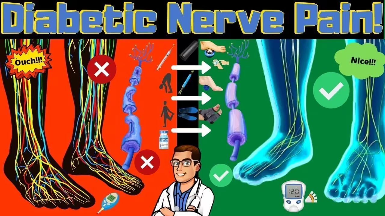
![Diabetes Peripheral Neuropathy Treatment [Diabetic Nerve Pain Remedy]](https://www.michiganfootdoctors.com/wp-content/cache/flying-press/83886306c119511909f4e50d00e25908.jpg)
Watch: Diabetes Peripheral Neuropathy Treatment [Diabetic Nerve Pain Remedy] — MichiganFootDoctors YouTube
OS1st FS4 — non-binding, moisture-wicking, protects fragile diabetic skin.
Recovery Slide for Indoor Wear
HOKA Ora 3 — protects diabetic feet from barefoot injury at home.
As an Amazon Associate, Balance Foot & Ankle earns from qualifying purchases. Product recommendations are based on clinical experience; prices and availability shown above update live from Amazon.
When to See a Podiatrist
One unnoticed blister on a neuropathic foot can become a limb-threatening ulcer in under 14 days. Medicare covers diabetic shoes (A5500) and comprehensive foot exams annually for most diabetic patients with neuropathy or circulation concerns. Balance Foot & Ankle runs a dedicated diabetic limb-preservation program — vascular screening, offloading, ulcer care, and shoe fitting — all in one visit. Schedule your annual diabetic foot exam today.
Call Balance Foot & Ankle: (810) 206-1402 · Book online · Offices in Howell & Bloomfield Township
Frequently Asked Questions About PAD and the Diabetic Foot
How do I know if I have PAD?
The most reliable way to screen for PAD is an ankle-brachial index (ABI) test — a painless, non-invasive comparison of blood pressure at the ankle and arm. All diabetic patients over 50 should have ABI screening, and those with additional risk factors (smoking, hypertension, dyslipidemia, family history) should be screened earlier. Symptoms like calf cramping with walking, foot coldness, slow-healing wounds, and skin changes on the feet may also indicate PAD. If you notice these signs, request a vascular evaluation at your next podiatry appointment.
Can PAD be reversed?
Atherosclerotic plaque cannot be fully reversed, but PAD progression can be significantly slowed and symptoms improved with aggressive risk factor management. Statin therapy, blood pressure control, smoking cessation, glycemic optimization (HbA1c below 7%), antiplatelet therapy (aspirin or clopidogrel), and supervised exercise programs all improve outcomes. Supervised walking exercise programs specifically have been shown to increase pain-free walking distance by 100-150% in claudication patients. These interventions, combined with protective foot care using proper insoles like PowerStep Pinnacle Maxx, represent the medical management foundation for all PAD patients.
How often should diabetic patients check their feet?
Every single day, without exception. Daily foot inspection is the most important self-care activity for diabetic patients, especially those with PAD. Check every surface of both feet — tops, bottoms, sides, between all toes, and around all toenails — looking for any change from the previous day. In addition to daily self-inspection, diabetic patients with PAD should see a podiatrist every 2-3 months for professional vascular assessment, nail care, and comprehensive foot examination.
What shoes are best for diabetic patients with PAD?
Diabetic PAD patients need shoes with extra depth to accommodate orthotics or thick insoles, firm yet cushioned soles, seamless interiors (no ridges or stitching that could create pressure points), and adjustable closures (laces or Velcro) to accommodate swelling fluctuations. Avoid pointed toes, high heels, and open-toed shoes. Medicare covers diabetic therapeutic shoes and custom insoles for qualifying patients — ask your podiatrist about the Therapeutic Shoe Program.
Can I use compression socks if I have diabetes and PAD?
It depends on your PAD severity. Patients with mild PAD (ABI above 0.8) can generally use graduated compression like DASS compression socks safely and may benefit from them if they also have venous insufficiency or dependent edema. Patients with moderate to severe PAD (ABI below 0.8) should not use standard compression without explicit clearance from their vascular specialist, as external compression can further compromise already-inadequate arterial blood flow. Always discuss compression with your treating physician before use.
Medical Sources & References
- Gerhard-Herman MD, Gornik HL, Barrett C, et al. 2016 AHA/ACC guideline on the management of patients with lower extremity peripheral artery disease. Circulation. 2017;135(12):e686-e725. doi:10.1161/CIR.0000000000000471
- Armstrong DG, Boulton AJM, Bus SA. Diabetic foot ulcers and their recurrence. N Engl J Med. 2017;376(24):2367-2375. doi:10.1056/NEJMra1615439
- Jude EB, Eleftheriadou I, Tentolouris N. Peripheral arterial disease in diabetes — a review. Diabet Med. 2010;27(1):4-14. doi:10.1111/j.1464-5491.2009.02866.x
- Mills JL Sr, Conte MS, Armstrong DG, et al. The Society for Vascular Surgery Lower Extremity Threatened Limb Classification System: risk stratification based on wound, ischemia, and foot infection (WIfI). J Vasc Surg. 2014;59(1):220-234. doi:10.1016/j.jvs.2013.08.003
- American Diabetes Association. Peripheral arterial disease in people with diabetes. Diabetes Care. 2003;26(12):3333-3341. doi:10.2337/diacare.26.12.3333
Watch: PAD and the Diabetic Foot Explained
Dr. Biernacki discusses peripheral arterial disease in diabetic patients, vascular assessment methods, and the limb salvage approach:
Diabetic? Get Your Vascular Foot Assessment
If you’re due for routine diabetic foot screening or have noticed changes in your foot’s appearance, sensation, or healing ability, Dr. Biernacki and the team at Balance Foot & Ankle provide comprehensive vascular assessments and coordinated care for diabetic patients with PAD throughout Southeast Michigan.
Related Diabetic Foot Resources
- Preventing Charcot Foot in Diabetic Neuropathy
- Diabetic Foot Osteomyelitis
- Ankle Swelling: Causes and Treatment
- Podiatrist Recommended Foot Care Products 2026
- Best Shoes for Plantar Fasciitis 2026
- Custom Orthotics in Southeast Michigan
Affiliate Disclosure: This page contains affiliate links to products we recommend. Balance Foot & Ankle may earn a small commission at no additional cost to you. We only recommend products Dr. Biernacki uses with patients in clinical practice.
Dr. Tom’s Recommended Products: See our clinically tested product recommendations for this condition. View Dr. Tom’s recommended products →
When to See a Podiatrist for PAD and Diabetic Foot Care
If you have diabetes with cramping in your calves when walking, non-healing foot wounds, or cold feet with absent pulses, you may have peripheral arterial disease. PAD dramatically increases the risk of amputation in diabetic patients. At Balance Foot & Ankle, we perform vascular assessments and coordinate comprehensive diabetic foot care at our Howell and Bloomfield Township offices.
→ Learn about our diabetic foot care services
→ Book your appointment
→ Call (810) 206-1402
Clinical References
- Marso SP, Hiatt WR. Peripheral arterial disease in patients with diabetes. J Am Coll Cardiol. 2006;47(5):921-929. doi:10.1016/j.jacc.2005.09.065
- Hingorani A, LaMuraglia GM, Henke P, et al. The management of diabetic foot: a clinical practice guideline by the Society for Vascular Surgery in collaboration with the American Podiatric Medical Association and the Society for Vascular Medicine. J Vasc Surg. 2016;63(2 Suppl):3S-21S.
- Jude EB, Eleftheriadou I, Tentolouris N. Peripheral arterial disease in diabetes — a review. Diabet Med. 2010;27(1):4-14. doi:10.1111/j.1464-5491.2009.02866.x
Insurance Accepted
BCBS · Medicare · Aetna · Cigna · United Healthcare · HAP · Priority Health · Humana · View All →
Howell Office
4330 E Grand River Ave
Howell, MI 48843
Get Directions →
Bloomfield Township Office
43494 Woodward Ave, Suite 208
Bloomfield Township, MI 48302
Get Directions →
Your Board-Certified Podiatrists



Ready to Get Back on Your Feet?
Same-week appointments available at both locations.
Book Your AppointmentIn Our Clinic
Diabetic neuropathy patients in our clinic often don’t realize they have it until we put a 10-gram Semmes-Weinstein monofilament to the plantar foot and they can’t feel it. Many arrive for an unrelated concern — an ingrown toenail, a callus — and we catch the neuropathy on screening. The conversation then shifts: we need to discuss daily foot inspections, appropriate footwear, the urgency of any blister or open area, and the timing of vascular referral if pulses are diminished. Comprehensive diabetic foot exams are covered by Medicare annually. If you have diabetes, we want to see you once a year even if nothing hurts.
In-Office Treatment at Balance Foot & Ankle
When conservative care isn’t enough, Dr. Tom Biernacki and the team at Balance Foot & Ankle offer advanced, same-day options — including Peripheral Arterial Disease (PAD) Foot Care in Michigan at our Howell and Bloomfield Township clinics.
Same-day appointments available. Call (810) 206-1402 or book online.
In-Office Treatment at Balance Foot & Ankle
If home treatment isn’t providing relief for your diabetic foot conditions, our podiatry team at Balance Foot & Ankle can help with same-day evaluations and advanced in-office care.
Same-day appointments available. (810) 206-1402
Frequently Asked Questions
What causes this condition?
Causes include mechanical stress, biomechanical imbalance, age-related changes, and sometimes systemic disease. Our clinical exam plus imaging identifies the specific driver.
Can it go away on its own?
Mild cases sometimes resolve with rest and supportive footwear. Persistent symptoms past 4-6 weeks rarely resolve without active treatment.
Is surgery required?
Most patients resolve with non-surgical care. Surgery is reserved for refractory cases or structural deformity.
What is Diabetic foot?
Diabetic foot is a common foot/ankle condition that affects mobility and quality of life. Understanding the underlying cause is the first step in successful treatment. Our podiatrists at Balance Foot & Ankle perform a hands-on biomechanical exam, review your activity history, and use diagnostic imaging when appropriate to identify the root cause—not just treat the symptom. Many patients have been told to “rest and ice” without a deeper diagnostic workup; our approach is different.
Symptoms and warning signs
Common signs of diabetic foot include pain that worsens with activity, morning stiffness, swelling, tenderness when palpated, and difficulty bearing weight. If you experience sudden severe pain, inability to walk, visible deformity, numbness or color change, contact our office the same day or visit urgent care—these can signal a more serious injury such as a fracture, tendon rupture, or vascular compromise. Diabetics with any foot wound should seek same-day care.
Conservative treatment options
Most cases of diabetic foot respond to non-surgical care: structured rest, supportive footwear changes, custom orthotics, targeted stretching and strengthening protocols, anti-inflammatory medications when medically appropriate, and in-office procedures such as ultrasound-guided injections. We also offer advanced therapies including MLS laser therapy, EPAT/shockwave, regenerative injections, and image-guided procedures. Treatment is sequenced from least invasive to most invasive, and we explain the rationale at every step.
When is surgery considered?
Surgery is reserved for cases that fail 3-6 months of well-structured conservative care, when there is structural pathology (severe deformity, complete tear, advanced arthritis), or when imaging shows damage that will not heal without intervention. Our surgeons have performed 3,000+ foot and ankle procedures and prioritize minimally-invasive techniques whenever appropriate. We discuss recovery timelines, return-to-activity milestones, and realistic outcome expectations before any procedure is scheduled.
American Diabetes Association: Diabetic Foot Care
Recovery timeline and prevention
Recovery from diabetic foot varies based on severity and chosen treatment path. Conservative cases often improve within 4-8 weeks with consistent adherence to the protocol. Post-procedural recovery may range from a few days (in-office procedures) to several months (reconstructive surgery). Long-term prevention involves footwear assessment, activity modification, structured strengthening, and regular check-ins with your podiatrist if you have a history of recurrence. We provide written home-exercise plans and digital follow-up support.
Ready to feel better?
Same-week appointments available in Howell and Bloomfield Township, Michigan.
Book Your Visit
Dr. Tom Biernacki, DPM is a board-certified foot & ankle surgeon (ABFAS & ABPM) at Balance Foot & Ankle Specialists in Southeast Michigan. With over a decade of clinical experience, he specializes in heel pain, bunions, diabetic foot care, sports injuries, and minimally invasive surgery. Dr. Biernacki is a member of the APMA and ACFAS, and his patient education content on MichiganFootDoctors.com and YouTube has made him one of the most-followed foot & ankle educators on YouTube.
More questions patients ask
How often should diabetics check their feet?
Diabetic patients should perform daily self-examinations of both feet, checking for cuts, blisters, redness, swelling, calluses, and temperature changes. Professional diabetic foot exams should occur at least annually, or every 3-6 months for patients with neuropathy, peripheral artery disease, or history of foot ulcers. Dr. Biernacki provides comprehensive diabetic foot care programs.
Why do diabetics have foot problems?
Diabetes causes peripheral neuropathy (nerve damage reducing sensation), peripheral artery disease (reduced blood flow), and impaired immune function. This combination means injuries go unnoticed, heal slowly, and are prone to infection. Approximately 15% of diabetics will develop a foot ulcer, and diabetic foot complications are the leading cause of non-traumatic amputation. Prevention through regular podiatric care is essential.
What are the signs of diabetic neuropathy in feet?
Early signs include tingling, burning, or numbness starting in the toes and progressing upward. Other symptoms include sensitivity to touch, muscle weakness, balance problems, and loss of reflexes. If you experience any of these symptoms, schedule an evaluation with Dr. Biernacki at 810-206-1402 for nerve conduction testing and a comprehensive neuropathy management plan.
Still have a question about coverage or cost? Book online and we will check your benefits before your visit: Book in Howell · Book in Bloomfield Township. Prefer to talk it through first? Call (810) 206-1402.