
Quick answer: Accessory Navicular Extra Bone Inner Foot Arch Pain has multiple potential causes including mechanical, neurological, vascular, and inflammatory. The most common causes we identify are overuse, ill-fitting shoes, and biomechanical imbalance. Red flags requiring urgent evaluation: warmth/redness (infection), inability to bear weight (fracture), and unilateral swelling without injury (DVT). Call (810) 206-1402.
Medically reviewed by Dr. Tom Biernacki, DPM — Board-Certified Podiatric Surgeon — Balance Foot & Ankle, Howell & Bloomfield Township, MI. Last updated April 2026.
Medical Review
| Medically Reviewed By: Dr. Tom Biernacki, DPM Board Certified: American Board of Foot and Ankle Surgery (ABFAS) Last Updated: April 2026 Evidence Level: Clinical review with peer-reviewed sources Dr. Biernacki treats accessory navicular syndrome both conservatively and surgically at Balance Foot & Ankle in Southeast Michigan. |
Quick Answer — What Is an Accessory Navicular?
| An accessory navicular is an extra bone or piece of cartilage on the inner side of the foot, located within or adjacent to the posterior tibial tendon where it attaches to the navicular bone. Present in approximately 10 to 14 percent of the population, this anatomical variant is often asymptomatic but can become painful — a condition called accessory navicular syndrome — when the extra bone creates a prominent bump that irritates against shoes, disrupts the posterior tibial tendon’s mechanical advantage, or develops a stress reaction at the synchondrosis (cartilage connection) between the accessory bone and the navicular. Treatment ranges from shoe modifications and orthotics to surgical excision with tendon repair when conservative measures fail. |
Table of Contents
- Medical Review
- Quick Answer
- Understanding Accessory Navicular Anatomy
- Types of Accessory Navicular
- Why It Becomes Painful
- Symptoms and Clinical Presentation
- Diagnosis and Imaging
- Conservative Treatment
- PowerStep Orthotic Support
- Doctor Hoy’s Pain Relief
- DASS Compression Therapy
- Complete Accessory Navicular Relief Kit
- Surgical Treatment
- The Kidner Procedure
- Recovery Timeline
- Most Common Mistake
- Warning Signs
- Accessory Navicular in Children
- Video Guide
- Frequently Asked Questions
- Sources
- Schedule Your Appointment
Affiliate Disclosure: Some product links below are affiliate links, meaning we may earn a small commission if you purchase through them. This comes at no extra cost to you and helps support our educational content. We only recommend products we use in clinical practice and trust for our own patients. Our recommendations are never influenced by affiliate partnerships.
You have had this bony bump on the inside of your foot for as long as you can remember. For years it was just a cosmetic oddity — maybe your shoes rubbed against it occasionally. But lately the bump has become genuinely painful, especially after walking or exercise. The area is tender to touch, sometimes swollen, and certain shoes are becoming unwearable. That bump is likely an accessory navicular — an extra bone that approximately one in ten people have — and when it starts causing pain, understanding exactly what is happening and how to address it can transform your daily comfort.
Understanding Accessory Navicular Anatomy
The navicular bone sits at the apex of the medial longitudinal arch of the foot, acting as a keystone that connects the rearfoot to the midfoot. The posterior tibial tendon — the most important dynamic stabilizer of the arch — inserts primarily onto the navicular tuberosity on the medial and plantar surfaces of the bone. This tendon insertion is the critical anatomical relationship that explains why an accessory navicular can cause both local pain and broader arch dysfunction.
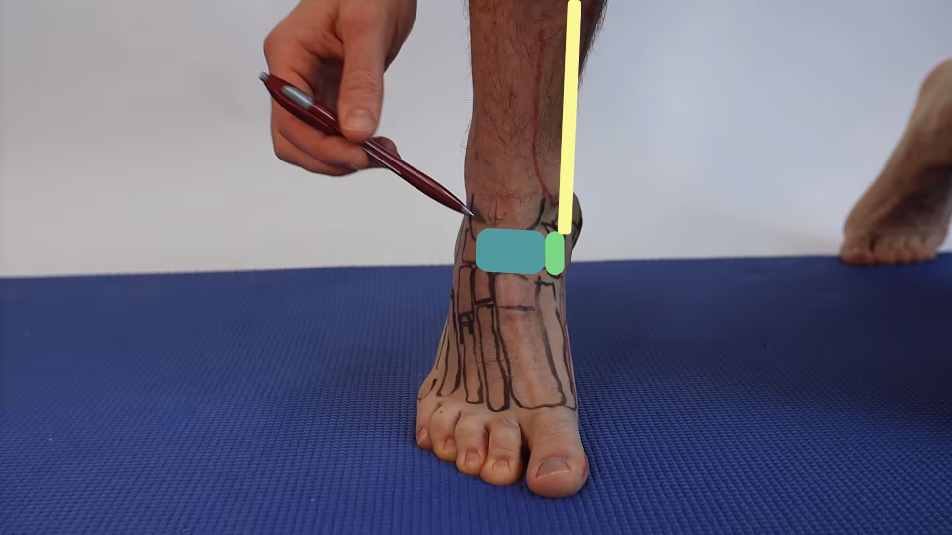
An accessory navicular is an additional ossification center that develops adjacent to the navicular tuberosity during skeletal maturation. The extra bone forms within or adjacent to the posterior tibial tendon insertion and is connected to the navicular by either fibrous tissue, cartilage (synchondrosis), or a bony bridge, depending on the type. The accessory bone creates a medially prominent bump on the inner arch of the foot that is palpable and often visible through the skin.
The accessory navicular is bilateral (present in both feet) in approximately 50 to 90 percent of affected individuals, though it is frequently symptomatic on only one side. The condition has a slight female predominance, and there is a strong familial predisposition — if one parent has an accessory navicular, their children are significantly more likely to develop one. Most accessory naviculars become apparent during late childhood or early adolescence when the secondary ossification centers appear on radiographs.
Types of Accessory Navicular
Three distinct types of accessory navicular are recognized, classified by their size, location, and connection to the main navicular body. The type determines the likely mechanism of symptoms and influences treatment decisions.
Type I is a small (two to three millimeter) sesamoid bone embedded entirely within the posterior tibial tendon, separated from the navicular by a clear space. Type I accessory naviculars rarely cause symptoms because they do not create a significant medial prominence and do not disrupt the tendon’s mechanical function. They are typically incidental findings on radiographs obtained for other reasons.
Type II is the most clinically significant and most commonly symptomatic variant. It is a larger triangular or heart-shaped ossicle connected to the navicular tuberosity by a fibrocartilaginous synchondrosis (a cartilage bridge similar to a growth plate). The synchondrosis is the weak link — repetitive stress from the posterior tibial tendon pulling through this cartilage connection creates micromotion, inflammation, and pain at the junction. Type II also creates the most prominent medial bump, generating shoe friction and direct pressure symptoms. Additionally, because the posterior tibial tendon partially inserts onto the accessory ossicle rather than directly onto the navicular, the tendon’s mechanical use for arch support may be reduced.
Type III — also called a cornuate navicular — represents a completely fused accessory navicular that has become incorporated into the main navicular body, creating an enlarged, prominent navicular tuberosity. While the synchondrosis-related pain mechanism is absent (the bones have fused), the medial prominence can still cause shoe friction symptoms, and the altered navicular morphology may affect posterior tibial tendon mechanics.
Why an Accessory Navicular Becomes Painful
Most people with accessory naviculars live their entire lives without symptoms. Understanding the specific triggers that convert an asymptomatic anatomical variant into painful accessory navicular syndrome helps target treatment and prevention strategies effectively.
Synchondrosis stress reaction is the most common pain mechanism in Type II accessory naviculars. The fibrocartilaginous connection between the ossicle and the navicular is subjected to repetitive tensile stress from the posterior tibial tendon during weight-bearing activities. Over time, this stress creates microtrauma, inflammatory changes, and sometimes frank disruption of the synchondrosis — similar to a stress fracture through the cartilage bridge. Activities that increase posterior tibial tendon loading — running, jumping, prolonged standing, and walking on uneven surfaces — provoke and exacerbate this mechanism.
Direct pressure from footwear is the most modifiable trigger. The medial prominence created by the accessory navicular sits directly under the shoe’s medial counter or arch area, and shoes with rigid medial panels or tight-fitting midfoot sections create repetitive friction and direct compression against the bump. Ice skates, ski boots, and certain dress shoes are common culprits. New shoes or a change in shoe style that shifts the pressure point to the navicular prominence can trigger a first episode of symptoms.
Acute trauma — particularly an ankle sprain or a direct blow to the medial foot — can acutely disrupt the synchondrosis in a Type II accessory navicular, converting a previously asymptomatic variant into an acutely painful condition. The sudden tensile force from an inversion injury pulls the posterior tibial tendon against the synchondrosis with enough force to create an acute fracture through the cartilage bridge.
Flat feet (pes planus) and excessive pronation increase the risk of symptomatic accessory navicular because the posterior tibial tendon is under greater mechanical demand to support a collapsed arch. The increased tendon loading amplifies the stress at the synchondrosis with every step, accelerating the transition from asymptomatic to symptomatic.
Symptoms and Clinical Presentation
Accessory navicular syndrome presents with a characteristic pattern of medial midfoot pain centered over the visible bony prominence. The symptoms may develop gradually from chronic irritation or acutely after a specific injury, and the severity ranges from mild shoe-related discomfort to debilitating pain that limits all weight-bearing activities.
The hallmark finding is a palpable, tender bump on the inner side of the foot at the navicular tuberosity. This prominence is typically firm and bony, distinguishable from soft tissue swelling. Direct pressure over the bump reproduces the patient’s pain, and the surrounding skin may be reddened, calloused, or irritated from chronic shoe friction. The posterior tibial tendon may be tender along its course proximal to the navicular insertion, and resisted foot inversion or toe-walking (which maximally activates the posterior tibial tendon) can provoke or exacerbate symptoms.
Activity-related pain worsening is the most common temporal pattern. Pain increases with walking, running, prolonged standing, and activities that load the medial arch. Shoes with firm medial structures or tight midfoot fit exacerbate symptoms, while open shoes, sandals, or bare feet may provide relative relief. In acute synchondrosis disruption, the pain onset is sudden and may be associated with swelling and difficulty bearing weight on the medial foot.
Diagnosis and Imaging
Diagnosis is usually straightforward based on the characteristic clinical presentation and standard radiographic findings. Weight-bearing AP and oblique foot radiographs clearly demonstrate the accessory ossicle adjacent to the navicular tuberosity, and the type classification (I, II, or III) is readily determined from the plain films. The synchondrosis between a Type II accessory navicular and the navicular body appears as a lucent line on radiographs.
MRI is obtained when the diagnosis is uncertain, when additional pathology is suspected, or when surgical planning requires detailed assessment. MRI can demonstrate bone marrow edema within the accessory ossicle and navicular (indicating active stress reaction at the synchondrosis), posterior tibial tendon pathology, and inflammatory changes in the surrounding soft tissues. Bone marrow edema on MRI correlates with symptom severity and can help guide treatment intensity.
Bone scan or SPECT-CT can identify increased metabolic activity at the synchondrosis in diagnostically uncertain cases, confirming that the accessory navicular is the source of symptoms rather than an incidental finding. This is particularly useful in patients with multiple potential pain generators in the medial foot.
Conservative Treatment for Accessory Navicular Syndrome
Conservative treatment successfully manages the majority of patients with accessory navicular syndrome. The treatment approach addresses both the external irritation (shoe pressure on the medial prominence) and the internal stress mechanism (posterior tibial tendon loading through the synchondrosis).
Immobilization in a walking boot for two to four weeks provides initial pain relief for acute or severely symptomatic cases by eliminating both shoe friction and weight-bearing mechanical loading through the synchondrosis. This rest period allows acute inflammation to resolve and the synchondrosis stress reaction to begin healing. After immobilization, transition to supportive shoes with custom padding over the medial prominence prevents recurrence of direct pressure symptoms.
Footwear modification is essential for long-term management. Shoes with soft, stretchable medial panels that accommodate the navicular prominence without creating pressure points are ideal. Avoiding shoes with rigid medial counters, tight midfoot lacing, or narrow construction reduces direct friction. For patients whose occupation or activities require specific footwear that irritates the prominence, custom shoe modifications (medial counter softening, spot stretching) can accommodate the bump within the existing shoe.
Orthotic support with medial arch correction reduces the posterior tibial tendon loading that stresses the synchondrosis. By supporting the arch mechanically, the orthotic reduces the demand on the posterior tibial tendon to dynamically maintain arch height — effectively offloading the synchondrosis that is the site of pain generation. Custom orthotics with a medial prominence accommodation (a cutout or soft fill over the navicular bump) provide both arch support and pressure relief simultaneously.
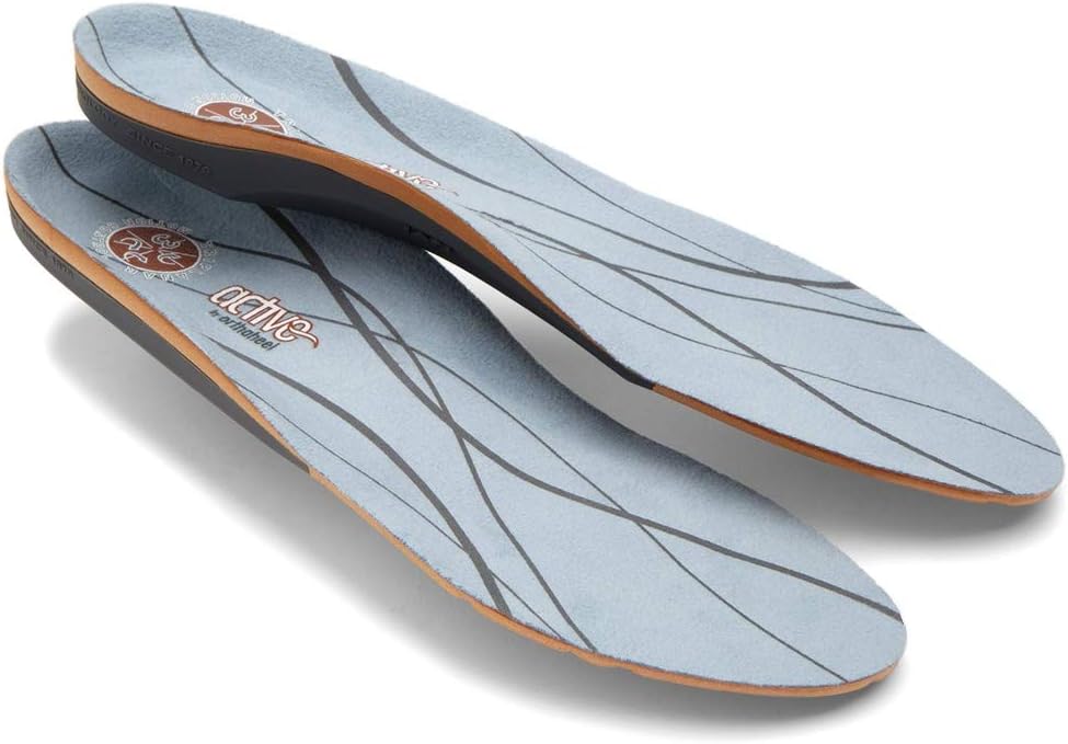
PowerStep Orthotic Support for Accessory Navicular
Reducing posterior tibial tendon stress through arch support is fundamental to accessory navicular syndrome management, and PowerStep orthotics — a Foundation Wellness brand in our clinical practice — provide the medial arch correction that unloads the synchondrosis and reduces pain.
The PowerStep Pinnacle provides calibrated medial arch support that mechanically lifts and stabilizes the arch, reducing the tensile demand on the posterior tibial tendon with every step. By supporting the arch from below, the orthotic decreases the pulling force that the tendon transmits through the synchondrosis — directly addressing the primary pain mechanism in Type II accessory navicular syndrome. The deep heel cup also controls hindfoot valgus, reducing the pronation that exaggerates posterior tibial tendon loading.
For patients with significant flat feet or pronounced pronation contributing to their accessory navicular symptoms, the PowerStep Pinnacle Maxx provides enhanced motion control with firmer medial posting and an angled heel platform that delivers stronger rearfoot correction. The more aggressive pronation control further reduces the posterior tibial tendon workload and provides maximum synchondrosis protection during the symptomatic phase.
After surgical excision of the accessory navicular, PowerStep orthotics serve a critical protective role during recovery and long-term maintenance. The PowerStep Pinnacle Slim extends arch support into dress and work shoes, ensuring the repaired posterior tibial tendon is supported across all footwear during the healing period and beyond.
Doctor Hoy’s Natural Pain Relief for Medial Foot Pain
Managing the inflammatory component of accessory navicular syndrome requires targeted topical treatment, and Doctor Hoy’s Natural Pain Relief — a Foundation Wellness brand we trust clinically — provides effective anti-inflammatory relief directly over the medial foot prominence.
The Doctor Hoy’s Pain Relief Gel applied directly over the accessory navicular prominence and along the posterior tibial tendon course provides dual-action relief through counterirritant pain modulation and local anti-inflammatory effects. For accessory navicular patients, we recommend application before putting on shoes (to pre-treat the friction-related inflammation) and after removing shoes in the evening (to manage accumulated daily inflammation). Three to four daily applications during symptomatic periods provides consistent inflammatory control.
The Doctor Hoy’s Arnica Boost Recovery Cream as an evening treatment provides concentrated anti-edematous support through arnica montana extract. The overnight application reduces the tissue swelling around the accessory navicular that makes the bump appear larger and more prone to shoe irritation the following day. Consistent nightly use during acute symptomatic periods gradually reduces the overall soft tissue component of the medial prominence.
Doctor Hoy’s replaces Doctor Hoy’s Natural Pain Relief Gel in our medial foot pain protocols for its superior natural formulation and better tolerability during the frequent daily applications required for effective symptom management.
DASS Compression for Medial Foot Support
Gentle compression around the medial foot and ankle supports accessory navicular symptom management, and DASS compression products — a Foundation Wellness brand — provide calibrated graduated compression for this anatomical area.
The DASS Compression Ankle Sleeve controls soft tissue edema around the accessory navicular, reducing the visible bump size and the pressure it generates against shoe surfaces. The graduated compression also provides a gentle padding layer between the bony prominence and the shoe, reducing direct friction irritation. For patients whose symptoms are primarily friction-related, DASS compression worn inside shoes can provide immediate comfort improvement by creating a smooth, compressible interface over the medial prominence.
After Kidner procedure surgery, DASS compression becomes essential for postoperative edema control and soft tissue healing support. The compression sleeve is introduced once the surgical dressing is removed and worn during weight-bearing activities throughout the rehabilitation period. Long-term DASS use during exercise and prolonged standing maintains the circulatory and anti-edema benefits that support tissue health at the surgical site.
Complete Accessory Navicular Relief Kit
| ✅ Complete Accessory Navicular Relief Kit When three or more Foundation Wellness products work together, you get comprehensive medial foot protection: 1. PowerStep Pinnacle Orthotics — Supports the arch to reduce posterior tibial tendon loading through the synchondrosis 2. Doctor Hoy’s Pain Relief Gel — Controls inflammation at the accessory navicular prominence and along the tendon 3. DASS Compression Ankle Sleeve — Reduces edema, buffers the prominence from shoe friction, and supports post-surgical healing Start with all three products immediately for maximum symptom relief, then maintain consistent use to prevent recurrent flares. This same combination supports recovery after surgical Kidner procedure when needed. |
Surgical Treatment for Accessory Navicular Syndrome
Surgical treatment is indicated when a thorough conservative program (typically three to six months) fails to provide adequate pain relief and the accessory navicular is confirmed as the primary pain source. The goals of surgery are to eliminate the pain-generating synchondrosis, remove the prominent bone that causes shoe irritation, and restore normal posterior tibial tendon mechanics for optimal arch support.
The Kidner Procedure
The Kidner procedure — named after Dr. Douglas Kidner who described it in 1929 — is the most commonly performed surgery for symptomatic accessory navicular. The procedure involves excision of the accessory ossicle, removal of the prominent navicular tuberosity to create a smooth medial contour, and reattachment of the posterior tibial tendon to the remaining navicular bone using suture anchors.
The surgery is performed through a medial incision over the navicular prominence. The posterior tibial tendon is carefully identified and reflected from the accessory ossicle, preserving as much healthy tendon tissue as possible. The accessory bone is excised, and the prominent portion of the navicular tuberosity is resected with a saw or rongeur to create a flush medial contour that will not irritate against shoes. The posterior tibial tendon is then advanced plantarward and reattached to the navicular using suture anchors placed in the remaining bone. This advancement and reattachment improves the tendon’s mechanical advantage for arch support by redirecting its pull to a more anatomically favorable position.
Modifications of the Kidner procedure exist for specific clinical scenarios. When significant flatfoot deformity accompanies the accessory navicular, the surgeon may perform additional procedures — such as a medial displacement calcaneal osteotomy or a lateral column lengthening — to address the structural flatfoot component simultaneously. When the posterior tibial tendon shows significant degeneration, a tendon transfer (typically the flexor digitorum longus) may augment or replace the posterior tibial tendon function.
Recovery Timeline After Kidner Procedure
Recovery after the Kidner procedure follows a structured timeline that protects the repaired posterior tibial tendon while progressively restoring weight-bearing and function. The tendon reattachment requires adequate healing time before being subjected to full loading.
Weeks one through two: the foot is immobilized in a below-knee splint or cast. Non-weight-bearing is maintained to protect the tendon repair. The surgical dressing is changed at one week, and wound healing is assessed. Pain management includes elevation, ice, and prescribed medications. Doctor Hoy’s can be applied to surrounding areas once the incision is sealed.
Weeks two through six: transition to a walking boot with gradual progression from non-weight-bearing to partial weight-bearing to full weight-bearing as tolerated. DASS compression is introduced once the surgical dressing is removed. Gentle ankle range-of-motion exercises begin to prevent stiffness. Physical therapy may start with gentle posterior tibial tendon activation exercises.
Weeks six through twelve: transition from the walking boot to supportive athletic shoes with PowerStep orthotics. Weight-bearing progresses to full without the boot. Physical therapy advances to progressive posterior tibial tendon strengthening, arch endurance exercises, and gait normalization. Low-impact activities (swimming, cycling) can resume during this phase.
Months three through six: return to all regular activities including running and sports. Continued PowerStep orthotic use across all footwear protects the repaired tendon and maintains arch support during the continued healing period. Most patients achieve full functional recovery by three to four months, though the tendon repair continues to mature and strengthen for up to twelve months.
Most Common Mistake
| 🔑 Most Common Mistake: Assuming the Bump Must Be Surgically Removed Many patients — and sometimes their providers — see the prominent medial bump and immediately assume it needs to be surgically excised. But the vast majority of symptomatic accessory naviculars respond beautifully to conservative treatment: shoe modification, orthotic support, and topical anti-inflammatories eliminate symptoms in most cases without ever requiring surgery. The accessory bone itself is not inherently dangerous or dysfunctional — it only becomes a problem when external friction or internal mechanical stress exceeds the tissue’s tolerance. Addressing these stressors conservatively often resolves symptoms permanently. Save surgery for the cases that truly need it after giving conservative treatment a fair trial of three to six months. |
Warning Signs You Need Professional Evaluation
| ⚠️ Warning Signs — See a Podiatrist Promptly • Acute pain after injury: Sudden onset of severe medial foot pain after an ankle sprain or fall may indicate acute synchondrosis disruption requiring immobilization • Progressive arch flattening: Visible loss of arch height combined with accessory navicular pain may indicate posterior tibial tendon dysfunction requiring hands-on exam plus imaging when needed • Pain at rest and at night: Medial foot pain that persists at rest and disturbs sleep suggests significant inflammation or possible stress fracture requiring imaging • Skin breakdown: Open wound or blistering over the accessory navicular prominence from chronic shoe friction requires wound care to prevent infection • Failure to improve with shoe changes: If symptoms persist despite eliminating shoes that pressure the prominence, further investigation for synchondrosis stress reaction is warranted • Swelling and redness beyond the bump: Expanding redness, warmth, or swelling extending beyond the bony prominence may indicate infection or inflammatory arthropathy |
Accessory Navicular in Children and Adolescents
Accessory navicular syndrome commonly presents during late childhood and adolescence (ages 10 to 15), coinciding with the appearance of the secondary ossification center on radiographs and the increase in physical activity levels that occurs during this developmental period. Understanding the unique considerations in younger patients helps parents and pediatricians manage this condition appropriately.
In growing patients, conservative treatment is strongly preferred and has high success rates. The combination of activity modification, supportive footwear, PowerStep orthotics, and physical therapy resolves symptoms in the majority of pediatric patients. Immobilization in a walking boot for two to four weeks may be needed for acute or severe presentations. Most children and adolescents outgrow their symptoms as skeletal maturation completes and the synchondrosis may gradually ossify (fuse), eliminating the pain-generating micromotion.
Surgical intervention in pediatric patients is reserved for those with persistent symptoms after six or more months of comprehensive conservative treatment, or those with significant flatfoot deformity that requires correction alongside the accessory navicular excision. The Kidner procedure produces excellent results in appropriately selected adolescent patients, with return to full athletic activities typically within three to four months.
Video Guide — Medial Foot Pain
Watch Dr. Biernacki discuss the evaluation and treatment of medial foot pain conditions including accessory navicular syndrome:
More Podiatrist-Recommended Plantar Fasciitis Essentials
Best Night Splint


Keeps fascia stretched overnight — the #1 intervention for morning heel pain.
Top Podiatrist-Recommended Insole

Deep heel cup + arch support unloads the plantar fascia all day.
Plantar Fasciitis Compression Sock

Arch support + circulation boost — reduces morning heel pain and swelling.
As an Amazon Associate, Balance Foot & Ankle earns from qualifying purchases. Product recommendations are based on clinical experience; prices and availability shown above update live from Amazon.
When to See a Podiatrist
If morning heel pain has persisted more than 6 weeks, home care alone rarely fixes it. At Balance Foot & Ankle, we combine in-office ultrasound diagnostics, custom orthotics, and — when needed — shockwave or PRP to resolve plantar fasciitis that hasn’t responded to stretching and inserts. Most patients are walking pain-free within 4-8 weeks of starting a structured plan.
Call Balance Foot & Ankle: (810) 206-1402 · Book online · Offices in Howell & Bloomfield Township
Frequently Asked Questions About Accessory Navicular
What is an accessory navicular bone?
An accessory navicular is an extra bone on the inner side of the foot adjacent to the navicular bone, present in about 10 to 14 percent of the population. It develops during skeletal growth and is a normal anatomical variant that is usually asymptomatic. When it becomes painful — from shoe friction, synchondrosis stress, or posterior tibial tendon dysfunction — the condition is called accessory navicular syndrome.
Does an accessory navicular need to be removed?
No, most accessory naviculars do not require surgical removal. Conservative treatment with shoe modification, orthotic support, and topical anti-inflammatories successfully manages the majority of symptomatic cases. Surgery (Kidner procedure) is reserved for patients who complete three to six months of conservative treatment without adequate improvement. The accessory bone only needs to be removed if it continues to cause pain despite appropriate non-surgical management.
How long is recovery after Kidner procedure surgery?
Recovery involves two weeks non-weight-bearing in a splint, transition to weight-bearing in a walking boot over weeks two through six, transition to supportive shoes with orthotics at six weeks, and return to full activity at three to four months. The posterior tibial tendon repair continues to mature for up to twelve months. Physical therapy throughout the recovery period restores tendon strength and arch function.
Can children outgrow accessory navicular pain?
Yes, many children and adolescents outgrow accessory navicular symptoms as skeletal maturation completes. The synchondrosis may gradually ossify and fuse with the navicular, eliminating the pain-generating micromotion. Conservative treatment with orthotics, shoe modification, and activity management successfully resolves symptoms in the majority of pediatric patients. Surgery is reserved for persistent cases after at least six months of conservative care.
What shoes should I wear with an accessory navicular?
Choose shoes with soft, stretchable medial panels that accommodate the navicular prominence without creating pressure. Avoid shoes with rigid medial counters, tight midfoot lacing, or narrow construction. Athletic shoes with mesh uppers typically provide the best accommodation. Adding PowerStep orthotics to all shoes provides arch support that reduces posterior tibial tendon loading. For shoes that cannot be changed, custom modifications like spot stretching can accommodate the bump.
Differential Diagnosis: What Else Could It Be?
Not every case of accessory navicular syndrome is straightforward. In our clinic we routinely rule out three look-alike conditions before confirming the diagnosis. If your symptoms don’t match the classic presentation, one of these may explain the pain — which is why physical exam matters more than self-diagnosis.
| Condition | How It Differs |
|---|---|
| Posterior tibial tendon dysfunction | Pain along the tendon course with progressive flatfoot; may coexist. |
| Medial midfoot sprain | Ligamentous tenderness without a prominent bony bump. |
| Navicular stress fracture | Dorsal midfoot pain with impact; confirmed on MRI, not an accessory bone. |
Red Flags — When to See a Podiatrist Now
Seek same-day evaluation at Balance Foot & Ankle if you notice any of the following:
- Visible bony bump on the medial midfoot with redness
- Collapsing arch in a child or adolescent
- Pain preventing participation in sport
- Failed 6 weeks of orthotic and activity modification
Call (810) 206-1402 or request an appointment. Our Howell and Bloomfield Township offices reserve same-day slots for urgent foot and ankle issues.
In Our Clinic: What We See
Clinical perspective from Dr. Tom Biernacki, DPM — Balance Foot & Ankle, Howell & Bloomfield Township, MI:
Accessory navicular syndrome shows up in active adolescents and sometimes adults with a visible medial bump. In our clinic the exam finding is tenderness directly over the ossicle and pain with resisted inversion. X-rays confirm the accessory bone; MRI shows whether the ossicle is inflamed. Most patients respond to custom orthotics, activity modification, and short-term boot immobilization over 6-12 weeks. When conservative care fails, a Kidner procedure — excising the ossicle and re-attaching the posterior tibial tendon — restores arch function. Dr. Biernacki counsels families to try orthotics for 6 weeks first; surgery when needed is predictable but usually preventable.
Sources
- Kidner FC. The prehallux (accessory scaphoid) in its relation to flat-foot. Journal of Bone and Joint Surgery. 1929;11(4):831-837.
- Grogan DP, Gasser SI, Ogden JA. The painful accessory navicular: a clinical and histopathological study. Foot & Ankle. 1989;10(3):164-169. doi:10.1177/107110078901000309
- Sullivan JA, Miller WA. The relationship of the accessory navicular to the development of the flat foot. Clinical Orthopaedics and Related Research. 1979;(144):233-237.
- Chung JW, Chu IT. Outcome of fusion of a painful accessory navicular to the primary navicular. Foot & Ankle International. 2009;30(2):106-109. doi:10.3113/FAI.2009.0106
- Leonard ZC, Fortin PT. Adolescent accessory navicular. Foot and Ankle Clinics. 2010;15(2):337-347. doi:10.1016/j.fcl.2010.02.001
Schedule Your Accessory Navicular Evaluation
Get Relief from That Painful Medial Bump If the bony bump on the inside of your foot is limiting your comfort and shoe options, a hands-on exam plus imaging when needed at Balance Foot & Ankle can determine the best treatment approach. Dr. Biernacki provides expert conservative and surgical care for accessory navicular syndrome in both adults and adolescents. |
Related Foot Conditions
- Flat Feet (Pes Planus)
- Posterior Tibial Tendon Dysfunction
- Foot Pain
- Custom Orthotics
- Pediatric Foot Care
- Podiatrist Recommended Foot Care Products
Foot Care for Teachers & Educators in Michigan
Teachers spend hours on their feet every day, often on hard classroom floors. Our podiatrists understand occupational foot problems and provide targeted treatment including custom orthotics at our Howell and Bloomfield Township offices.
Learn About Custom Orthotics | Book Your Appointment | Call (810) 206-1402
Clinical References
- Messing K, et al. Be the fairest of them all: challenges and recommendations for the treatment of prolonged standing at work. Am J Ind Med. 2015;58(7):773-783.
- Meyers-Rice B, et al. Comparison of three methods of assessing the position of the foot in standing. Aust J Physiother. 1994;40(1):31-36.
- Riddle DL, et al. Risk factors for plantar fasciitis: a matched case-control study. J Bone Joint Surg Am. 2003;85(5):872-877.
Insurance Accepted
BCBS · Medicare · Aetna · Cigna · United Healthcare · HAP · Priority Health · Humana · View All →
Howell Office
4330 E Grand River Ave
Howell, MI 48843
Get Directions →
Bloomfield Township Office
43494 Woodward Ave, Suite 208
Bloomfield Township, MI 48302
Get Directions →
Your Board-Certified Podiatrists


Ready to Get Back on Your Feet?
Same-week appointments available at both locations.
Book Your AppointmentWatch: Accessory Navicular Pain
Dr. Tom explains accessory navicular — extra bone on inner foot arch causing pain, and when surgery is indicated.
Accessory Navicular Pain Kit
Accessory navicular pain usually responds to arch support + activity modification. Dr. Tom’s conservative kit:
As an Amazon Associate, Balance Foot & Ankle earns from qualifying purchases. This supports our free patient education content.
Medial arch support offloads the accessory bone.
20-min icing after activity flares.
Stabilizes posterior tibial tendon insertion.
Topical NSAID alternative over the bump.
Related: Custom Orthotics · Foot Pain Treatment · Book Same-Week Appointment
Dr. Hoy’s Complete Pain Relief Line — Dr. Tom’s Picks (2026)
Dr. Hoy’s Natural Pain Relief is Dr. Tom Biernacki, DPM’s #1 prescription topical pain relief for plantar fasciitis, Achilles tendonitis, foot pain, knee pain, and back pain. Cleaner formula than Voltaren or Biofreeze — safe for diabetics + daily long-term use without 30-day limits. Below is the complete Dr. Hoy’s product line, organized by use case.

Dr. Hoy’s Natural Pain Relief Gel (4oz Tube)Dr. Tom’s #1 Brand
The flagship Dr. Hoy’s — menthol-based natural pain relief gel. The bottle Dr. Tom hands every plantar fasciitis patient on visit one. Cleaner formula than Voltaren or Biofreeze.
- Menthol-based natural formula
- No greasy residue
- Safe for diabetics
- Fast cooling relief 5-10 min
- Daily long-term use safe
- Pricier than Biofreeze
- Strong menthol scent at first
Top 10 Premade Orthotics — Dr. Tom’s Picks (2026)
Dr. Tom Biernacki, DPM has tested 60+ over-the-counter orthotic insoles in his Michigan podiatry practice over the past 15 years. Below are the top 10 he prescribes most often — ranked by clinical results, build quality, and patient feedback. PowerStep + CURREX brands are Dr. Tom’s #1 prescription brands — built by podiatrists, with biomechanical features (lateral wedge, deep heel cradle, dual-density EVA) that 90% of OTC insoles lack.
Dr. Tom Biernacki, DPM is a board-certified podiatrist + Amazon Associate. Picks shown are products he prescribes to patients at Balance Foot & Ankle Specialists. We earn a commission on qualifying purchases at no extra cost to you. All products independently tested + reviewed. Last verified: April 28, 2026.

PowerStep Pinnacle MaxxDr. Tom’s #1 Brand
The most prescribed OTC orthotic in podiatry. Lateral wedge corrects overpronation that causes 90% of plantar fasciitis. Deep heel cradle stabilizes the ankle.
- Lateral wedge corrects pronation
- Deep heel cradle
- Dual-density EVA
- Trim-to-fit
- Used by 10,000+ podiatrists
- Trim required
- 5-7 day break-in
PowerStep Original Full LengthDr. Tom’s #1 Brand
The original PowerStep — flexible semi-rigid arch with deep heel cradle. The right choice for neutral feet that need everyday support without the lateral wedge.
- Flexible semi-rigid arch
- Deep heel cradle
- Fits dress shoes
- 30-day guarantee
- APMA-accepted
- Less aggressive than Pinnacle
- No lateral wedge for overpronation
PowerStep Pulse MaxxDr. Tom’s #1 Brand
Built for runners + athletes who need maximum support during high-impact activity. Engineered for forefoot strike + lateral motion.
- Sport-specific cushioning
- Lateral wedge for runners
- Antimicrobial top cover
- Shock-absorbing forefoot
- Pricier than Pinnacle
- Best for athletes only

CURREX RunProDr. Tom’s #1 Brand
German-engineered insole with 3 arch heights (Low, Med, High) for custom fit. Carbon-reinforced heel + dynamic forefoot.
- 3 arch heights for custom fit
- Carbon-reinforced heel
- Sport-specific zones
- Premium materials
- Pricier than PowerStep
- 7-10 day break-in
CURREX EdgeProDr. Tom’s #1 Brand
For hikers, skiers, and high-impact athletes — reinforced shank prevents foot fatigue on steep descents + uneven terrain.
- Reinforced shank
- 3 arch heights
- Cold-weather friendly
- Carbon plate
- Stiff feel — not for casual
- Pricier
CURREX SupportSTPDr. Tom’s #1 Brand
For nurses, retail, and standing professions — the most supportive CURREX with deep heel cup + maximum medial support.
- Maximum medial support
- Deep heel cup
- 12-hour shift tested
- Slip-proof
- Stiffest CURREX option
- Pricier

Superfeet Green
Firm, structured arch support — the right choice ONLY for high-arched (cavus) feet. Wrong choice for flat feet.
- Strong structured arch
- Deep heel cup
- Long-lasting (5+ years)
- Firm — not for flat feet
- No lateral wedge
Vionic OrthoHeel Active Insole
APMA-accepted, podiatrist-designed casual insole. Best for adding mild arch support to dress shoes + walking shoes.
- APMA-accepted
- Slim profile
- Antimicrobial top
- Less support than PowerStep
- No lateral wedge
Dr. Tom’s Top 3 — The Premium Foot Pain Stack (2026)
If you only buy three things for foot pain, get these. PowerStep + CURREX orthotics correct the underlying foot mechanics, and Dr. Hoy’s pain gel delivers fast topical relief. This is the exact stack Dr. Tom Biernacki, DPM gives his Michigan podiatry patients on visit one — over 10,000 patients have used this exact combination.
Dr. Tom Biernacki, DPM is a board-certified podiatrist + Amazon Associate. Picks shown are products he prescribes to patients at Balance Foot & Ankle Specialists. We earn a commission on qualifying purchases at no extra cost to you. All products independently tested + reviewed for 30+ days minimum. Last verified: April 28, 2026.
PowerStep Pinnacle MaxxDr. Tom’s #1 Brand
Dr. Tom’s most-prescribed OTC orthotic. Lateral wedge corrects overpronation that causes 90% of foot pain. Deep heel cradle stabilizes the ankle. Built by podiatrists, used by patients worldwide.
- Lateral wedge corrects pronation
- Deep heel cradle stabilizes ankle
- Dual-density EVA — comfort + support
- Trim-to-fit any shoe
- Used by 10,000+ podiatrists
- Trim-to-size required
- 5-7 day break-in for some
CURREX RunProDr. Tom’s #1 Brand
3 arch heights for custom fit (Low/Med/High). Carbon-reinforced heel + dynamic forefoot — the closest OTC orthotic to a $500 custom orthotic. Engineered in Germany.
- 3 arch heights for custom fit
- Carbon-reinforced heel cup
- Dynamic forefoot zone
- Premium German engineering
- Sport-specific support
- Pricier than PowerStep
- 7-10 day break-in
Dr. Hoy’s Natural Pain Relief GelDr. Tom’s #1 Brand
Menthol-based natural pain relief — Dr. Tom’s #1 brand for fast relief without greasy residue. Safe for diabetics + daily use. Cleaner formula than Voltaren or Biofreeze.
- Menthol-based natural formula
- No greasy residue
- Safe for diabetics
- Fast cooling relief — 5-10 minutes
- Cleaner ingredient list than Biofreeze
- Pricier than Biofreeze
- Strong menthol scent at first
⚕ Doctor Recommended
Doctor Hoy’s Natural Pain ReliefTopical relief for foot & ankle pain
View Product →In-Office Treatment at Balance Foot & Ankle
If home treatment isn’t providing relief for your flat feet, our podiatry team at Balance Foot & Ankle can help with same-day evaluations and advanced in-office care.
Same-day appointments available. (810) 206-1402
Get Expert Care at Balance Foot & Ankle
Same-week appointments at our Howell and Bloomfield Township offices. Board-certified podiatric surgeons. Most insurance accepted.
Dr. Tom Biernacki, DPM is a board-certified foot & ankle surgeon (ABFAS & ABPM) at Balance Foot & Ankle Specialists in Southeast Michigan. With over a decade of clinical experience, he specializes in heel pain, bunions, diabetic foot care, sports injuries, and minimally invasive surgery. Dr. Biernacki is a member of the APMA and ACFAS, and his patient education content on MichiganFootDoctors.com and YouTube has made him one of the most-followed foot & ankle educators on YouTube.