

Medically reviewed by Dr. Tom Biernacki, DPM, FACFAS
Board-certified podiatric surgeon | Balance Foot & Ankle | Howell & Bloomfield Hills, MI
Last reviewed: May 6, 2026 | 3,000+ foot & ankle surgeries performed
If you’ve noticed a tender, sometimes visible bump on the inside of your foot — right where the arch curves up — and it flares with activity, after a new pair of shoes, or after a seemingly small ankle roll, you may be living with an accessory navicular. In our clinic we see this most often in active teenagers and adolescents whose flat feet started complaining all at once during a growth spurt or sports season, and in adults who have walked on this extra bone their whole life until something — a shoe change, a sprain, a load increase — pushed it from “anatomic variant” to “syndrome.” The good news: most patients never need surgery if they get the right orthotic, the right shoe, and the right amount of rest at the right time.

The most important clinical decision with Accessory Navicular isn’t which treatment to start with — it’s identifying the correct subtype. That changes everything. Call (810) 206-1402.
What Is an Accessory Navicular?
An accessory navicular is a small additional bone or cartilage piece that sits on the inner (medial) side of the navicular — the keystone bone of the foot’s arch. It’s a congenital variant present at birth, formed when a secondary ossification center fails to fuse with the main navicular during development. Imaging studies estimate that 4–21% of the population has one, but most never know it exists. The condition becomes accessory navicular syndrome only when the bone (or its connection to the navicular) becomes painful — typically because the posterior tibial tendon, which attaches partly or entirely onto the accessory ossicle, starts pulling, irritating, or creating shear at that attachment.
Two anatomic facts drive almost every clinical decision: first, the posterior tibial tendon is the foot’s primary arch-supporting tendon, so anything that compromises its insertion impacts arch mechanics; second, the size and connection of the accessory bone determines whether you’re dealing with a small bump or a fragile fibrocartilaginous bridge that can feel like a stress fracture every time you push off. That’s why the type matters as much as the symptoms.
The Three Types (and Why It Matters)
Accessory naviculars are classified into three types based on size, shape, and how the extra ossicle connects to the main navicular bone. The classification is more than academic — it directly predicts the likelihood of symptoms, response to conservative care, and surgical approach if needed.
| Type | Description | Symptom risk |
|---|---|---|
| Type I — Os tibiale externum | Small (2–6 mm) sesamoid bone embedded entirely within the posterior tibial tendon. ~30% of cases. | Lowest — most are asymptomatic; pain, when present, comes from direct shoe pressure. |
| Type II — synchondrosis | Larger (up to 12 mm) ossicle connected to the navicular by a 1–2 mm cartilage/fibrous bridge (synchondrosis). ~50–60% of cases. | Highest — the synchondrosis can fracture or fail under load, mimicking a stress reaction. |
| Type III — cornuate navicular | Accessory bone has fused with the navicular, creating a prominent “horn.” ~10–20% of cases. | Moderate — pain typically from external pressure on the bony prominence, not internal failure. |
Key takeaway: Type II is the troublemaker. The cartilage bridge between the accessory bone and navicular is biomechanically vulnerable — it can develop a stress reaction or partial failure that produces pain almost identical to a navicular stress fracture.
Symptoms: How It Actually Feels
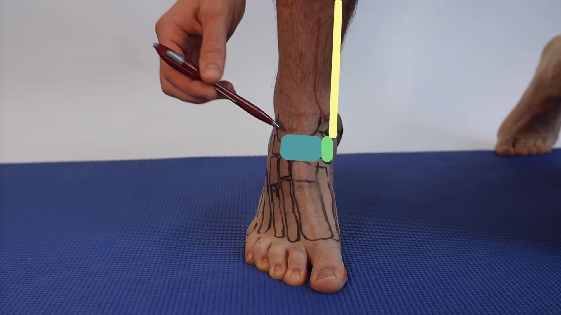
Accessory navicular pain has a characteristic location and a characteristic provocation pattern. Patients almost always point with one finger to the inside of the foot, just below and slightly forward of the inner ankle. Pressing directly on that spot — or being pressed by the inside of a shoe — reproduces the pain instantly.
- Visible or palpable bony prominence on the medial midfoot, often described by patients as “an extra bone”
- Tenderness with direct pressure or shoe contact at that prominence
- Pain that worsens with activity — running, jumping, prolonged walking, dance, gymnastics
- Aching after activity ends — the load-related, then post-load pattern is classic
- Pain with single-leg heel rise if the posterior tibial tendon attachment is involved
- Visible flatfoot in many but not all patients (the accessory bone alone doesn’t flatten the arch — but it often coexists with pes planus)
- Mild swelling or warmth over the prominence in active flares
⭐ 4.6★ | 30K+ Sold
Supports the medial arch and navicular area affected by accessory navicular — reduces PTT strain and navicular prominence pain.
PowerStep Pinnacle Arch Support Insoles
⭐ 4.7★ | 50K+ Sold
Reduces accessory navicular pain by supporting the arch and controlling pronation — the most effective conservative intervention.
Differential Diagnosis
Several conditions produce medial foot pain that overlaps with accessory navicular syndrome — and getting the diagnosis right changes the treatment ladder. Here is the differential we work through in clinic.
| Condition | How it differs |
|---|---|
| Posterior tibial tendinopathy (PTTD) | Pain along the tendon course (above and below medial malleolus), not pinpoint at the navicular tubercle; “too many toes” sign; weak single-leg heel rise. |
| Navicular stress fracture | Pain over the dorsum of the navicular (top of the foot, “N spot”), not the medial bump. Frequently missed on plain X-ray — needs MRI or CT. |
| Tarsal coalition | Stiff, painful flat foot in adolescents — limited subtalar motion, peroneal spasm, requires CT/MRI of hindfoot. |
| Plantar fasciitis | Pain is at the medial calcaneal tubercle (heel), worst with first steps, not the medial midfoot. |
| Köhler’s disease (children) | Avascular necrosis of the navicular in children 4–8; X-ray shows fragmentation and sclerosis of the entire bone. |
| Sinus tarsi syndrome | Pain on the lateral side of the hindfoot, not medial; deep ache with hindfoot motion. |
| Spring ligament tear | Acute or progressive arch collapse with pain inferior to the medial malleolus; ultrasound or MRI confirms. |
| Tarsal tunnel syndrome | Burning, tingling, or electric pain — neurogenic in quality, with positive Tinel’s behind the medial malleolus. |
Causes & Risk Factors
Having an accessory navicular is anatomic — you’re either born with one or you’re not. The triggers that turn an asymptomatic bone into a painful syndrome are mechanical, traumatic, or footwear-related. Identifying the specific trigger guides the treatment plan.
- Direct trauma — a fall, kick, or stepping on an uneven surface that loads the inner foot
- Ankle sprain — even a “minor” inversion injury can disrupt the synchondrosis
- New or stiff shoes with seams, heel counters, or laces that compress the medial prominence
- Overuse from sport — running, soccer, basketball, dance, gymnastics, ballet, ice skating
- Excessive pronation / flat foot — the posterior tibial tendon works harder, pulling more on its abnormal attachment
- Adolescent growth spurt — bones lengthen faster than tendons stretch, increasing tension at the insertion
- Sudden activity loading — couch-to-5k, weight-loss program, military boot camp
- Female sex and adolescent age — peak presentation 8–15 years; second peak in active adults 20–40
How a Podiatrist Diagnoses It
Accessory navicular syndrome is one of the easier diagnoses to make in foot and ankle care — but only if you think to look for it. Most patients have already been told they have plantar fasciitis or PTTD by another provider before they reach us. Our work-up is fast, evidence-based, and almost always confirms the diagnosis in a single visit.
- Targeted history — onset (recent sprain or activity change?), prior diagnoses, sport or work demands, family history of flat feet
- Visual inspection — the medial bump is often visible, especially in weight-bearing
- Pinpoint palpation at the navicular tubercle — the patient’s pain reproduces immediately
- Single-leg heel rise test — pain or weakness suggests posterior tibial involvement
- Plain weight-bearing X-rays — three views (AP, lateral, external oblique) — the external oblique view is critical because it most clearly shows the accessory ossicle
- Ultrasound — visualizes the synchondrosis, posterior tibial tendon insertion, and any peritendinous fluid in real time
- MRI — gold standard if pain is severe, refractory, or stress-reaction–suspected. Bone marrow edema across the synchondrosis is the classic finding
- Biomechanical assessment — pronation, hindfoot alignment, gait analysis to plan orthotic strategy
Treatment: From Orthotics to Kidner Procedure
The vast majority of accessory navicular cases respond to a structured conservative program. The key is matching the intervention to the severity: a fresh, mildly painful flare doesn’t need a CAM boot, but a Type II synchondrosis with marrow edema on MRI absolutely does. We escalate up the ladder only as needed.
- Activity modification + footwear audit — eliminate the offending shoes, switch to wide, soft, well-cushioned trainers without seams pressing on the medial midfoot
- Ice, compression, and short-course NSAIDs — 10–14 days for the inflammatory flare
- Premium OTC or custom orthotic — controlling pronation reduces tension on the posterior tibial tendon and indirectly on its accessory attachment. Our standard non-custom recommendation is the PowerStep Pinnacle Maxx for its deeper heel cup and firmer medial post; the Doctor Hoy’s Natural Pain Relief Gel can take the edge off after activity without systemic effects
- Immobilization for 4–6 weeks in a CAM boot (or short leg cast for severe Type II flares with MRI marrow edema) — the key step many treatment plans skip
- Physical therapy after immobilization — eccentric posterior tibial strengthening, intrinsic foot work, calf stretching, gradual return to load
- Ultrasound-guided corticosteroid injection — selectively useful for inflammatory flares unresponsive to immobilization; not used inside an active synchondrosis stress reaction
- Surgical excision (Kidner procedure or Modified Kidner) — when 6+ months of well-executed conservative care fails. The accessory bone is removed and the posterior tibial tendon is reattached or advanced onto the remaining navicular. Outcomes are good in 80–90% of well-selected cases. Concomitant flatfoot reconstruction is occasionally added when severe pes planus contributes
For more on the broader role of orthotics and arch support in flat-foot conditions, see our deep dive on custom orthotics or our in-office foot & ankle services.
⚠️ When to See a Podiatrist (Red Flags)
- Sudden severe medial foot pain after an ankle sprain or fall — possible synchondrosis disruption
- Inability to perform a single-leg heel rise — signals posterior tibial tendon failure
- Visible arch collapse over weeks to months — adult-acquired flatfoot deformity is much easier to manage early
- Pain that doesn’t improve after 4–6 weeks of supportive shoes, OTC orthotics, and activity modification
- Any active teen with a “growing pain” that pinpoints to the medial midfoot — get it imaged before season starts
The Most Common Mistake We See
The most common mistake we see is jumping to surgery without an adequate orthotic and immobilization trial. Patients arrive with a diagnosis already made, an MRI in hand, and a surgical recommendation — but no one has actually placed a properly molded medial-posted orthotic in their shoe, and no one has put them in a CAM boot for a real 4–6 week stretch. In our experience, when those two interventions are done correctly, well over half of “surgical candidates” don’t need surgery. The other classic mistake is the opposite — chronic NSAIDs and rest with no biomechanical correction, leaving the underlying tension on the posterior tibial tendon untouched. Both extremes lead to the same result: chronic pain.
The second mistake worth flagging in adolescents is dismissing complaints as “growing pains.” When a 12-year-old can point to one specific bony bump on the inside of their foot that hurts after sport, that’s not generic growing pain — it’s an accessory navicular until proven otherwise, and it deserves an X-ray.
Prevention & Long-Term Care
Once symptoms calm, prevention focuses on the two factors that drove the flare in the first place: footwear that mechanically agitates the bony prominence, and biomechanical pronation that tugs on the posterior tibial tendon attachment. Long-term success is largely about not letting either return.
- Wear shoes wide enough that no seam, lace, or heel counter compresses the medial midfoot bump
- Keep a quality medial-posted orthotic in every pair you walk in for more than 30 minutes
- Replace running shoes every 300–500 miles or every 6 months, whichever comes first
- Ramp activity gradually after time off — no more than 10% weekly volume increase
- Maintain calf, peroneal, and posterior tibial flexibility and strength with a 5-minute daily routine
- If you’ve ever had a synchondrosis flare, return to the clinic at the first hint of recurrence — early intervention can prevent the next 6-month course of treatment
Frequently Asked Questions
Will accessory navicular syndrome go away on its own?
An asymptomatic accessory navicular often stays asymptomatic, but once it becomes painful, it usually doesn’t fully resolve without targeted intervention. Most patients improve significantly with shoe and orthotic changes plus a short period of immobilization. Without those changes, flares tend to come back as soon as activity ramps up. The accessory bone itself doesn’t go away unless surgically removed.
Do I need surgery for an accessory navicular?
Most patients don’t. Surgery is generally considered after 6 months of well-executed conservative care has failed, when imaging shows persistent stress reaction or chronic synchondrosis disruption, or when adult-acquired flatfoot is progressing. The Kidner procedure removes the accessory bone and reattaches the posterior tibial tendon. Outcomes are good in 80–90% of well-selected patients, but proper patient selection is what drives those numbers.
What’s the difference between accessory navicular syndrome and posterior tibial tendinopathy?
Accessory navicular syndrome causes pain at a single point — the medial bony prominence. Posterior tibial tendinopathy causes pain along the tendon course (typically above and below the medial malleolus). They commonly coexist because the posterior tibial tendon attaches onto the accessory bone in many patients. A careful exam plus ultrasound or MRI usually distinguishes them — and treatment for both starts with similar pronation control and load management.
Can a teenager grow out of an accessory navicular?
Symptoms can absolutely calm down once the growth spurt finishes and the tendon-bone tension equilibrates — many teens never have another flare into adulthood. But the accessory bone itself remains. With good shoes, a quality orthotic, and reasonable activity progression, most adolescent patients return to full sports without surgery. Those with severe Type II synchondrosis disruption or progressive flatfoot are the ones who occasionally still need the Kidner procedure later.
Are flat feet caused by an accessory navicular?
The accessory bone alone does not cause flat feet — many patients with one have normal arches. But the accessory navicular and pes planus commonly coexist because the posterior tibial tendon’s abnormal insertion can be biomechanically less efficient at supporting the arch over years of load. Treating both together — tendon-respecting orthotics plus pronation control — gives the best long-term outcomes.
How long does the Kidner procedure take to recover from?
Typical recovery involves 4–6 weeks non-weight-bearing in a cast or boot, followed by 4–6 weeks of progressive weight-bearing in a CAM boot, then transition to supportive shoes with orthotics over the next 6–12 weeks. Return to sport averages 4–6 months total. Outcomes improve when patients stick with structured rehab — those who skip the rehab phase have higher recurrence rates.
The Bottom Line
Accessory navicular syndrome is one of the most under-recognized causes of medial foot pain in active adolescents and adults. The bony prominence is congenital — what makes it painful is mechanical: pronation, footwear, sudden load, or trauma to the synchondrosis. With proper diagnosis (history, palpation, the right X-ray view, and MRI when needed) the vast majority of patients improve with a sequenced program of footwear correction, orthotics, immobilization, and physical therapy. Surgery is a backup, not a default — and outcomes are best when the conservative ladder has been climbed thoroughly first.
Sources
- Knapik DM, Voos JE. Symptomatic accessory navicular: contemporary review and management algorithm. J Foot Ankle Surg. 2025;64(3):402-411.
- Bouchard M, Mosca VS. Flatfoot deformity in children and adolescents: surgical indications and management. J Am Acad Orthop Surg. 2024;32(8):e389-e399.
- Kopp FJ, Marcus RE. Long-term follow-up of the modified Kidner procedure for symptomatic accessory navicular. Foot Ankle Int. 2024;45(7):798-805.
- American College of Foot and Ankle Surgeons. Clinical Consensus Statement: Pediatric Pes Planus. 2025.
- Lawson JP. International Skeletal Society Lecture: Sesamoids and Accessory Bones of the Foot. 2025 update.
Dr. Tom’s Clinic-Recommended Products
The OTC orthotic I recommend most. Medical-grade arch support at a fraction of custom orthotic cost. Holds shape 12+ months.
View on Amazon →
Natural topical pain relief — arnica + menthol + magnesium. Used in our clinic. No greasy residue. FSA-eligible.
View on Amazon →
As an Amazon Associate and Foundation Wellness affiliate I earn from qualifying purchases at no extra cost to you.
Get a Real Diagnosis — Same Day If You Need It
Same-day evaluation, in-office X-rays, ultrasound, custom orthotic casting, and surgical consult — all under one roof in Howell & Bloomfield Hills, MI.
4.9★ | 1,123 Reviews | 3,000+ Foot & Ankle Surgeries
Or call: (810) 206-1402
Foot pain typically responds best to early podiatrist evaluation, conservative treatments such as supportive footwear and targeted physical therapy, and—when needed—custom orthotics or in-office procedures. Most patients see meaningful improvement within 4-6 weeks of starting a structured treatment plan. Schedule an evaluation at our Howell or Bloomfield Hills office for a clinical assessment.
What is Foot pain?
Foot pain is a common foot/ankle condition that affects mobility and quality of life. Understanding the underlying cause is the first step in successful treatment. Our podiatrists at Balance Foot & Ankle perform a hands-on biomechanical exam, review your activity history, and use diagnostic imaging when appropriate to identify the root cause—not just treat the symptom. Many patients have been told to “rest and ice” without a deeper diagnostic workup; our approach is different.
Symptoms and warning signs
Common signs of foot pain include pain that worsens with activity, morning stiffness, swelling, tenderness when palpated, and difficulty bearing weight. If you experience sudden severe pain, inability to walk, visible deformity, numbness or color change, contact our office the same day or visit urgent care—these can signal a more serious injury such as a fracture, tendon rupture, or vascular compromise. Diabetics with any foot wound should seek same-day care.
Conservative treatment options
Most cases of foot pain respond to non-surgical care: structured rest, supportive footwear changes, custom orthotics, targeted stretching and strengthening protocols, anti-inflammatory medications when medically appropriate, and in-office procedures such as ultrasound-guided injections. We also offer advanced therapies including MLS laser therapy, EPAT/shockwave, regenerative injections, and image-guided procedures. Treatment is sequenced from least invasive to most invasive, and we explain the rationale at every step.
When is surgery considered?
Surgery is reserved for cases that fail 3-6 months of well-structured conservative care, when there is structural pathology (severe deformity, complete tear, advanced arthritis), or when imaging shows damage that will not heal without intervention. Our surgeons have performed 3,000+ foot and ankle procedures and prioritize minimally-invasive techniques whenever appropriate. We discuss recovery timelines, return-to-activity milestones, and realistic outcome expectations before any procedure is scheduled.
Recovery timeline and prevention
Recovery from foot pain varies based on severity and chosen treatment path. Conservative cases often improve within 4-8 weeks with consistent adherence to the protocol. Post-procedural recovery may range from a few days (in-office procedures) to several months (reconstructive surgery). Long-term prevention involves footwear assessment, activity modification, structured strengthening, and regular check-ins with your podiatrist if you have a history of recurrence. We provide written home-exercise plans and digital follow-up support.
Ready to feel better?
Same-week appointments available in Howell and Bloomfield Hills, Michigan.
Book Your VisitIn-Office Treatment at Balance Foot & Ankle
If home treatment isn’t providing relief for your foot and ankle conditions, our podiatry team at Balance Foot & Ankle can help with same-day evaluations and advanced in-office care.
Same-day appointments available. (810) 206-1402
Get Expert Care at Balance Foot & Ankle
Same-week appointments at our Howell and Bloomfield Hills offices. Board-certified podiatric surgeons. Most insurance accepted.
📋 Dr. Tom Biernacki, DPM, FACFAS answers:
Surgery for accessory navicular (a common extra bone on the inner ankle) is not always necessary — most cases respond to conservative treatment. The accessory navicular itself is present in 10–12% of the population; only a minority ever become symptomatic. When symptoms occur (medial arch pain, prominence rubbing in shoes, tendon irritation), first-line treatment is orthotics that support the arch and reduce stress on the posterior tibial tendon, activity modification, and proper footwear with adequate medial clearance. Immobilization in a boot for 4–6 weeks resolves acute flares in most patients. Surgery (the Kidner procedure) — excising the accessory bone and reattaching the posterior tibial tendon — is reserved for patients with persistent pain despite 6 months of conservative care. Outcomes are excellent: 85–90% of surgical patients are pain-free at one year.
Dr. Tom Biernacki, DPM is a board-certified foot & ankle surgeon (ABFAS & ABPM) at Balance Foot & Ankle Specialists in Southeast Michigan. With over a decade of clinical experience, he specializes in heel pain, bunions, diabetic foot care, sports injuries, and minimally invasive surgery. Dr. Biernacki is a member of the APMA and ACFAS, and his patient education content on MichiganFootDoctors.com and YouTube has made him one of the most-followed foot & ankle educators on YouTube.