
Quick answer: Bumps on the feet have different causes by location: bunions and bone spurs (bony), ganglion cysts or plantar fibromas (soft lumps), and warts or corns (skin). Most are harmless, but a bump that grows, hurts, changes color, or appears suddenly should be checked by a podiatrist.
Patients routinely misidentify the type of foot bump they have — and the most common misdiagnosis isn’t random. The exact location of a bump on the foot (sole, side, top, between toes, at a joint) is the single most predictive variable separating lesions that need surgical excision from those that resolve on their own. Call (810) 206-1402 — expert podiatric care across Michigan.
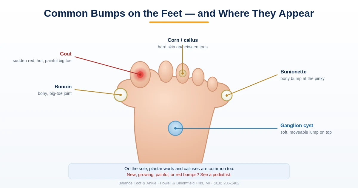
Table of Contents
Medically reviewed by Dr. Tom Biernacki, DPM
Board-certified podiatric surgeon | Balance Foot & Ankle
Last reviewed: May 2026
- Where Is the Bump? Quick Location Guide
- Red, Itchy & Pimple-Like Bumps
- Corns & Calluses
- Plantar Warts
- Bunions
- Ganglion Cysts
- Other Causes
- How to Tell Them Apart
- Treatment Overview
- When to See a Doctor
- FAQ
Bumps on the feet are among the most common foot complaints we see at Balance Foot & Ankle. Most people notice them when they start causing pain in certain shoes, or when they catch a glimpse in the mirror and realize something looks different. The good news: bumps on the feet are rarely serious. The better news: most respond well to treatment once properly identified.
The challenge is that different types of bumps look similar at first glance but require entirely different treatment approaches. A plantar wart treated like a callus won’t go away. A ganglion cyst treated like a wart may actually resolve spontaneously. Getting the diagnosis right is step one.
Where Is the Bump? Quick Location Guide
Location is the fastest way to narrow down what a foot bump is. Find where yours is, then jump to that section for details and treatment.
| Location | Most Likely Causes | Typical Character |
|---|---|---|
| Bottom of foot | Plantar wart, callus, plantar fibroma | Hard or rough; fibromas are firm knots in the arch |
| Top of foot | Ganglion cyst, bone spur (exostosis) | Soft and movable (cyst) or hard and fixed (spur) |
| Base of big toe | Bunion (hallux valgus) | Hard bony angle, worse in narrow shoes |
| Base of pinky toe | Tailor’s bunion (bunionette) | Smaller mirror-image of a bunion |
| Back of heel | Haglund’s deformity (“pump bump”) | Firm bony ridge aggravated by rigid heel counters |
| Tops of toes / between toes | Corns, hammertoe friction | Hard cap of thickened skin over pressure points |
One red flag overrides location: any bump that grows quickly, feels hard and fixed, or aches at night needs prompt evaluation — see the FAQ below.
Corns and Calluses
Corns and calluses are the most common bumps on the feet — areas of thickened, hardened skin that develop in response to repeated friction or pressure. The foot is particularly prone to them because it bears the entire body’s weight and experiences constant friction from footwear.
A callus is a broad, flat area of thickened skin typically found on the ball of the foot or heel. It usually doesn’t have a hard central core. A corn is a smaller, more focal area of thickening with a hard, cone-shaped core that points inward — this central nucleus is what causes the sharp, stabbing pain when you walk on it. Hard corns typically form on the tops of toes or on the outer surface of the little toe. Soft corns form between toes where moisture softens them.
The root cause is almost always mechanical: footwear that’s too tight, too narrow, or improperly fitted, combined with foot structure that concentrates pressure in certain spots. Custom orthotics can redistribute pressure and dramatically reduce corn and callus recurrence in patients with structural contributors.
The Most Common Mistake With Bumps on the Feet
The most common mistake patients make is self-treating a foot bump without knowing what it is. Rubbing a corn pad on a tailor’s bunion, using salicylic acid on a ganglion cyst, or trying to “pop” a plantar wart all delay proper care and can cause skin damage. The fix: location and texture tell you almost everything. A firm, bony bump on the outer foot is a tailor’s bunion. A soft, moveable bump is almost always a ganglion cyst. A rough, cauliflower-textured bump on the bottom of the foot is a plantar wart. When in doubt, a 15-minute office visit gives you a definitive answer and a targeted treatment plan.
Plantar Warts
Plantar warts are caused by human papillomavirus (HPV) — specifically strains 1, 2, 4, 60, and 63 that infect the skin. They enter through tiny cuts or breaks in the skin on the bottom of the foot, typically in moist public environments like pools, locker rooms, and gym showers.
Plantar warts look like calluses but have two distinguishing features: they interrupt the normal skin ridge lines (dermatoglyphics) — calluses don’t — and they contain tiny black dots (“seeds”) that are actually thrombosed capillaries, not seeds. Pinching a plantar wart side-to-side causes pain; pushing down on it directly causes less pain. This is the opposite of a callus, which hurts most with direct downward pressure.
Plantar warts can be solitary or occur in clusters (mosaic warts). They’re more common in children and immunocompromised individuals but can affect anyone. Treatment options include salicylic acid, cryotherapy (freezing), laser ablation, or surgical excision. Untreated warts can persist for years and spread.
Bunions
A bunion is a bony prominence at the base of the big toe, visible as a bump on the inner side of the foot. It’s not a growth of new bone — it’s a structural misalignment of the first metatarsophalangeal (MTP) joint where the big toe angles toward the second toe, causing the metatarsal head to protrude. Bunions are primarily hereditary, with footwear accelerating symptoms but not causing the deformity de novo.

- REDUCES SHOE FRICTION and PRESSURE
- IMMEDIATE, ALL-DAY PAIN RELIEF
- STAYS ON ALL DAY & NIGHT
- FITS EASILY in shoes
- Water & sweat resistant
Bunions are a major source of forefoot pain, particularly in women who wear narrower shoes. The bump itself can become inflamed (bursitis over the bunion), and the misalignment creates secondary hammertoes, corns, and metatarsalgia. Surgery — a bunionectomy or osteotomy — is the only definitive correction; conservative care (wide shoes, orthotics, toe spacers) manages symptoms but doesn’t reverse the deformity.
Ganglion Cysts and Other Cysts
A ganglion cyst is a fluid-filled sac arising from a joint capsule or tendon sheath. On the foot, they most commonly appear on the top of the foot or around the ankle but can form anywhere. They feel soft and slightly rubbery — distinctly different from the hard firmness of a bunion or bone spur. A key test: they transmit light (transillumination positive) in a dark room.
Ganglion cysts can appear suddenly, vary in size day to day, and may disappear spontaneously. They’re benign but can cause pain if they press on a nerve or make shoe fit difficult. Aspiration (draining with a needle) provides relief with a 50% recurrence rate; surgical excision has a lower recurrence rate but carries standard surgical risks.
Other Causes of Bumps on the Feet
Additional causes we see regularly include:
- Blisters — fluid-filled bumps from acute friction or heat. Usually self-resolving; should not be popped (especially in diabetic patients).
- Cysts (epidermoid/sebaceous) — keratin-filled lumps under the skin. Soft, moveable, may have a small pore on the surface. Can become infected and require drainage.
- Plantar fibromatosis (Ledderhose disease) — firm fibrous nodules embedded in the plantar fascia. More common in middle-aged adults; can cause arch pain.
- Lipoma — benign fatty tumor. Soft, moveable, non-tender. Can occur on the foot but more common elsewhere.
- Haglund’s deformity — a bony bump on the back of the heel where the Achilles tendon attaches. Sometimes called “pump bump” because it’s aggravated by rigid-backed shoes (like pumps).
- Tailor’s bunion (bunionette) — a bony prominence at the base of the fifth (pinky) toe. Mirror image of a standard bunion on the outer side of the foot.
Red, Itchy, or Pimple-Like Bumps
Not every foot bump is structural. Clusters of small red or fluid-filled bumps are usually a skin condition rather than a bone or soft-tissue mass:
- Athlete’s foot (tinea pedis) — itchy, scaly, sometimes blistering patches, classically between the toes or along the arch border. Often on both feet.
- Dyshidrotic eczema — crops of small, intensely itchy fluid-filled bumps on the soles and the sides of the toes; flares with heat, stress, and sweating.
- Friction blisters and heat rash — appear after long days, new shoes, or hot conditions, and settle within days once the trigger is removed.
- Insect bites — isolated red itchy bumps, usually on exposed skin.
The pattern matters: red and itchy points to skin and inflammation; hard, fixed, and slowly growing points to the structural causes covered above. Spreading redness, drainage, fever, or any new bump on a diabetic foot warrants a same-week appointment. More skin-focused guides are in our nail & skin conditions hub.
How to Tell the Bumps Apart at Home
Here’s a practical guide to help identify the most common foot bumps before your appointment:
| Characteristic | Corn/Callus | Plantar Wart | Bunion | Ganglion Cyst |
|---|---|---|---|---|
| Texture | Hard, rough skin | Rough, with black dots | Hard bony protrusion | Soft, rubbery |
| Pain type | Direct pressure | Side-to-side squeeze | At joint, in shoes | Variable; may be none |
| Location | Pressure points | Bottom of foot | Base of big toe | Top/inner foot |
| Skin ridges | Preserved | Interrupted | N/A (bony) | N/A (under skin) |
| Moves under skin? | No | No | No (fixed bone) | Yes (partially) |
Treatment Overview
The right treatment depends entirely on the correct diagnosis. Treating a plantar wart with corn pads won’t work. Here’s a high-level treatment guide:
- Corns & calluses — pumice stone or gentle filing after soaking, cushioning pads to relieve pressure, wider shoes, custom orthotics for structural contributors. Podiatric debridement for large or painful lesions.
- Plantar warts — over-the-counter salicylic acid (40%) for 8–12 weeks; professional cryotherapy, laser, or surgical removal for resistant warts.
- Bunions — wide toe-box shoes, bunion pads, toe spacers, orthotics. Surgery (osteotomy, Lapiplasty) when conservative care fails.
- Ganglion cysts — observation for asymptomatic cysts; aspiration or surgical excision for symptomatic ones.
⚠️ See a podiatrist if:
- Any bump is rapidly growing or changing in appearance
- Pain is severe enough to cause limping or change your gait
- You have diabetes or peripheral vascular disease — even “minor” bumps carry higher infection risk
- An open sore, bleeding, or ulceration develops over any bump
- Over-the-counter treatments have failed after 8 weeks
Learn more about how custom orthotics work — our Michigan guide covers casting, materials, activity-specific designs, and what to expect from the fitting process.
Footwear & Orthotics for Pressure-Related Foot Bumps
Bumps from friction or pressure often improve with better-fitting shoes. See our podiatrist-recommended shoes and recommended orthotics to offload the area, and book an evaluation if a bump changes or hurts.
In-Office Evaluation at Balance Foot & Ankle
A foot bump that grows, changes shape, or causes persistent pain needs clinical evaluation to rule out serious causes. At Balance Foot & Ankle, Dr. Tom Biernacki DPM provides bunion treatment and hammertoe correction for patients in Howell and Bloomfield Hills, MI.
Same-day appointments available. (810) 206-1402
Not sure what the bump is? Any new or growing lump on the foot is worth having looked at. A foot & ankle specialist can identify it and rule out anything serious — Balance Foot & Ankle sees patients same-week in Howell and Bloomfield Hills, MI.

Specialist For This Condition
Dr. Carl Jay, DPM is the Balance Foot & Ankle reconstructive surgeon other doctors refer to when persistent bumps, masses, or growths on the foot need surgical assessment. Fellowship-trained in complex foot and ankle reconstruction. Call (810) 206-1402 to request at the Howell or Bloomfield Hills office.
Frequently Asked Questions
Are bumps on the bottom of the foot serious?
Most bumps on the bottom of the foot are benign — plantar warts, calluses, and plantar fibromatosis are the most common causes. They can be painful but aren’t dangerous in otherwise healthy adults. The exception is in patients with diabetes or poor circulation, where any skin break or pressure lesion carries a higher risk of infection and slow healing. Any bump that doesn’t have an obvious benign explanation, is growing rapidly, or is associated with systemic symptoms warrants evaluation.
What causes small bumps on the toes?
Small bumps on the toes are most commonly corns (hard or soft), blisters, or warts. Hard corns typically form on the tops of the lesser toes where they rub against shoes. Soft corns form in the web spaces between toes where moisture traps them. Warts can form on the toes but are more common on the plantar surface. If you notice small flesh-colored bumps specifically on the toe knuckles in a person with rheumatoid arthritis, these could be rheumatoid nodules.
Can bumps on the feet go away without treatment?
It depends on the type. Blisters almost always resolve on their own within 1–2 weeks. Ganglion cysts frequently disappear spontaneously (and then recur). Plantar warts can resolve without treatment in children within 2 years in about 60–70% of cases; adult warts are more persistent. Corns and calluses persist and worsen as long as the mechanical cause continues. Bunions do not resolve without intervention.
What causes a hard lump on the bottom of the foot?
The most common hard lumps on the plantar surface are plantar fibromas (benign fibrous growths in the plantar fascia), plantar warts (verrucae), and foreign-body granulomas. Less commonly, a ganglion cyst, accessory ossicle, or malignant tumor may present this way. A podiatrist can distinguish these by location, mobility, and ultrasound if needed.
Are bumps on top of the foot serious?
Bumps on the dorsum (top) of the foot are most often ganglion cysts, extensor tendon ganglia, or bony exostoses — all typically benign. A bump that grows rapidly, is hard and fixed, or causes night pain warrants prompt evaluation to rule out bone tumors, which are rare but do occur in the foot.
When should I see a doctor for a foot lump?
See a podiatrist if: the lump is growing rapidly, is painfully firm, limits walking, bleeds, or has been present more than 4 weeks without explanation. Diabetic patients should seek evaluation for any new foot lump regardless of symptoms, as infection and Charcot bone changes can present as painless swellings.
The Bottom Line
Most bumps on the feet are benign and manageable. The key is correct identification: a callus needs different treatment than a wart, which needs different treatment than a bunion. When in doubt — or when a bump is painful, growing, or not responding to home treatment — a podiatry visit provides a quick answer and a clear treatment path.
Sources
- Loo SK, Tang WY. “Warts (non-genital).” Clinical Evidence. 2014;2014:1710.
- Menz HB, Lord SR. “Foot problems, functional impairment, and falls in older people.” Journal of the American Podiatric Medical Association. 1999;89(9):458-467.
- Singh D, Bentley G, Trevino SG. “Callosities, corns, and calluses.” BMJ. 1996;312(7043):1403-1406.
- Hecht PJ, Lin TJ. “Hallux valgus.” Medical Clinics of North America. 2014;98(2):227-232.
- American Academy of Orthopaedic Surgeons: Bunions — OrthoInfo
- American Academy of Dermatology: Warts — AAD.org
Related guide: The most common firm foot lump is a plantar fibroma. See Plantar Fibroma Treatment — causes, progression, and the conservative-to-surgical treatment ladder.
Related foot-skin guides
More on bumps and skin changes on the feet:
Ready to Get Relief?
Same-day appointments available in Howell & Bloomfield Hills, MI
4.9★ | 1,123 Reviews | 3,000+ Surgeries
Or call: (810) 206-1402
Dr. Tom Biernacki, DPM is a board-certified foot & ankle surgeon (ABFAS & ABPM) at Balance Foot & Ankle Specialists in Southeast Michigan. With over a decade of clinical experience, he specializes in heel pain, bunions, diabetic foot care, sports injuries, and minimally invasive surgery. Dr. Biernacki is a member of the APMA and ACFAS, and his patient education content on MichiganFootDoctors.com and YouTube has made him one of the most-followed foot & ankle educators on YouTube.