
You are in the right place. Dr. Tom Biernacki, DPM, FACFAS — board-certified foot & ankle surgeon with 3,000+ surgeries — explains exactly what bunionette / tailor’s bunion means and what actually works. Call (810) 206-1402 for a same-day appointment at our Howell or Bloomfield Township office.
The most important clinical decision with Bunionette isn’t which treatment to start with — it’s identifying the correct subtype. That changes everything. Call (810) 206-1402.
Related Conditions
Quick answer: A bunionette (tailor’s bunion) is a bony prominence at the base of the fifth toe caused by a widened fifth metatarsal head or metatarsus quintus varus. Most cases resolve with wide toe-box shoes, padding, and orthotics. Surgery (osteotomy) is reserved for cases unresponsive to 3–6 months of conservative care and produces excellent long-term results in 90%+ of patients.
Board-Certified Podiatric Foot & Ankle Surgeon · Last reviewed: May 5, 2026

Medically reviewed by Dr. Tom Biernacki, DPM
Board-certified podiatric surgeon | Balance Foot & Ankle
Last reviewed: April 2026
If you have a painful bump on the outside of your foot near your little toe that seems to get angrier with every pair of shoes you wear, you are likely dealing with a bunionette — also called a tailor’s bunion. It’s the fifth metatarsal’s answer to the classic bunion at the big toe, and while it gets far less attention, it can be just as disabling for the people who have it.
At Balance Foot & Ankle in Howell and Bloomfield Township, Michigan, Dr. Tom Biernacki treats bunionettes regularly using the full spectrum of interventions, from conservative management to minimally invasive surgical correction. Here is everything you need to know about this often-overlooked condition.
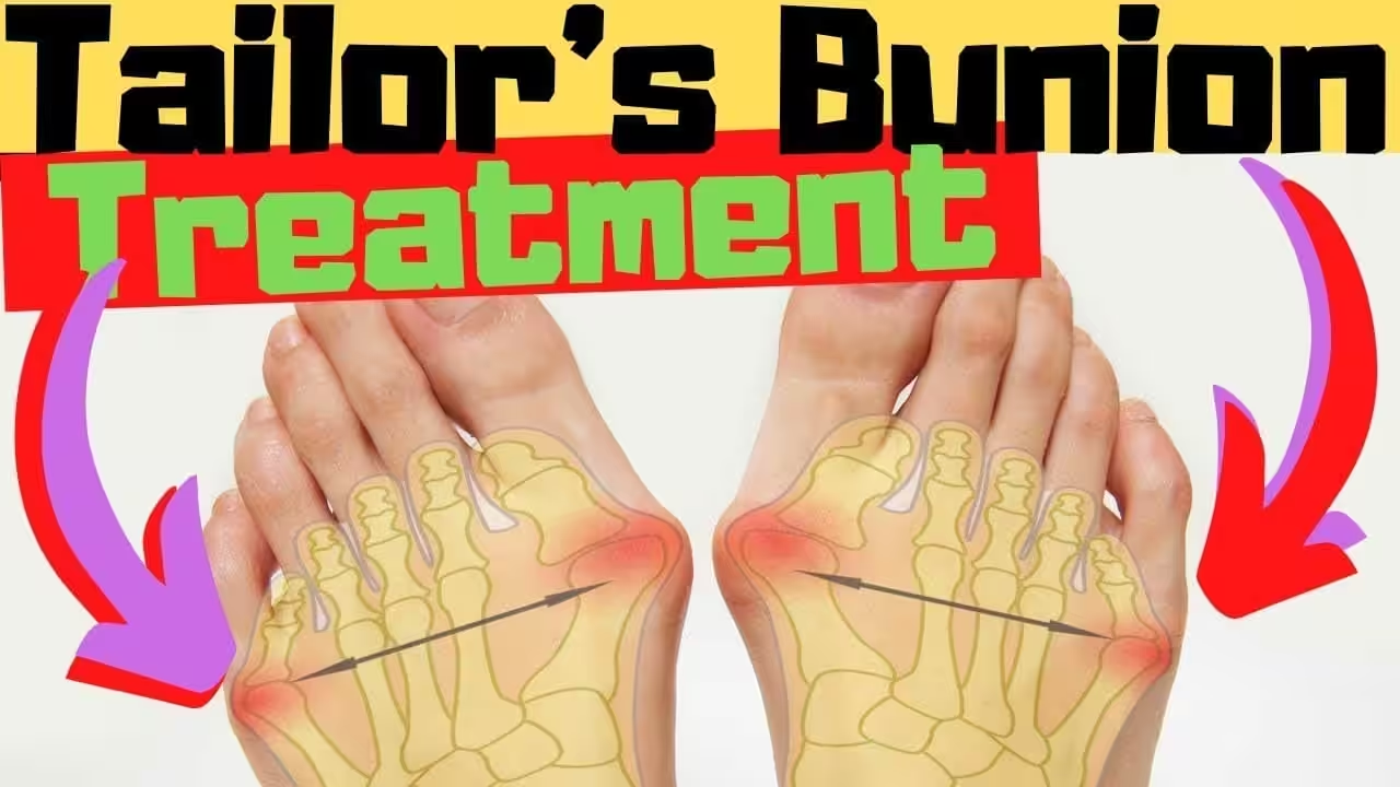
Table of Contents
- What Is a Bunionette (Tailor’s Bunion)?
- Symptoms
- Causes and Risk Factors
- Three Types of Bunionette Deformity
- Diagnosis
- Conservative Treatment
- Bunionette Surgery
- Frequently Asked Questions
What Is a Bunionette (Tailor’s Bunion)?
A bunionette, or tailor’s bunion, is a lateral prominence at the fifth metatarsophalangeal (MTP) joint — the joint where the little toe meets the fifth metatarsal bone. The name “tailor’s bunion” is historical: medieval tailors sat cross-legged for long hours, creating persistent pressure on the outer edge of the foot that caused this deformity to develop over time.
Unlike the classic bunion at the first MTP joint (which involves the big toe drifting toward the second toe), a bunionette involves the fifth metatarsal head flaring outward laterally, or the little toe drifting inward (toward the fourth toe), or a combination of both. The result is a prominent bony bump on the outer side of the forefoot that rubs against shoe walls, causing pain, callus formation, and bursitis.
Bunionettes are significantly less common than hallux valgus (first toe bunions) but are far more common than most people realize — affecting approximately 5-10% of people with symptomatic foot problems who present to podiatric care. In our clinic, we see them most frequently in women in their 40s–60s who wear narrow, pointed dress shoes.
Key takeaway: A bunionette is a structural deformity — the bone is actually splaying outward. This is why padding and toe separators provide temporary relief but cannot correct the underlying problem. Permanent correction requires surgical realignment of the fifth metatarsal.
Symptoms of a Bunionette
Bunionette symptoms are almost always directly related to shoe gear and worsen progressively as the deformity increases over years. Here is the typical presentation we see in our clinic:
- Visible bony prominence on the outer side of the foot at the base of the little toe — the most obvious finding
- Pain over the fifth metatarsal head — directly at the bump; sharp or aching; worse in shoes, better barefoot
- Shoe irritation — the bump rubs against shoe uppers; almost any closed-toe shoe causes discomfort
- Redness and swelling over the prominence — reactive bursitis developing under the skin over the fifth MTP joint
- Callus or corn formation over the lateral fifth metatarsal head — the skin thickens in response to chronic pressure
- Little toe deviation — the fifth toe drifts medially (toward the fourth toe), sometimes causing it to overlap or underlap
- Difficulty finding comfortable footwear — a hallmark complaint that brings patients to our clinic
Causes and Risk Factors for Bunionettes
Bunionettes develop from a combination of inherited foot structure and extrinsic mechanical forces — primarily from footwear. Here are the most important contributing factors:
- Hereditary foot morphology — a wide forefoot, splayed fifth metatarsal, or specific fifth metatarsal angulation is largely genetically determined. If a parent has a bunionette, the likelihood of developing one is significantly increased
- Narrow or pointed-toe footwear — the primary extrinsic driver. Shoes that compress the forefoot laterally push the fifth metatarsal head outward and the little toe inward over years
- High heels — transfer weight to the forefoot and compress the toes, accelerating deformity development
- Forefoot hypermobility — excessive motion at the fifth tarsometatarsal joint allows the fifth metatarsal to splay outward
- Flat feet — altered biomechanics change the pressure distribution across the forefoot, increasing lateral metatarsal loading
- Rheumatoid arthritis — joint destruction and synovitis can cause lateral deviation at any MTP joint, including the fifth
Three Types of Bunionette Deformity
The Coughlin classification system identifies three distinct types of bunionette based on the radiographic deformity pattern. Understanding the type is essential for surgical planning:
- Type 1 (most common — 50%+): Enlarged fifth metatarsal head — the metatarsal shaft is straight but the head itself is widened or has a lateral flange. Treatment: simple lateral condylectomy (shaving the lateral head prominence)
- Type 2 (lateral bowing): The fifth metatarsal shaft bows laterally through its entire length — a curved bone that pushes the head outward. Treatment: metatarsal shaft osteotomy to straighten the bone
- Type 3 (increased 4-5 intermetatarsal angle): The angle between the fourth and fifth metatarsals is abnormally large, causing the fifth metatarsal head to be positioned too far laterally. Treatment: proximal or distal metatarsal osteotomy to reduce the IM angle
In our clinic, we obtain weight-bearing X-rays for every bunionette patient to measure the 4-5 intermetatarsal angle and classify the deformity type. This classification directly determines which surgical approach produces the best outcomes — which is why “one-size-fits-all” surgical approaches for bunionettes consistently underperform.
Conservative Treatment for Bunionettes
The vast majority of bunionette patients achieve adequate symptom control with well-chosen conservative measures. Conservative treatment does not correct the underlying deformity, but it effectively manages the pain and inflammation that the deformity causes. Here is our evidence-based conservative protocol:
Footwear Modification
This is the single most effective conservative intervention. Shoes with a wide toe box, soft and flexible uppers, and a rounded (not pointed) toe shape eliminate the mechanical irritation that causes pain. We look for: toe box wide enough to accommodate the fifth metatarsal prominence without contact, soft leather or mesh uppers that can be stretched if needed, and low heel height to minimize forefoot loading. In our clinic, we can stretch specific shoe areas over a last to create a “bunionette window” that accommodates the prominence in existing footwear.
Padding and Protective Cushioning
Donut-shaped gel or foam pads placed over the bunionette redistribute pressure away from the prominence — providing immediate pain relief that is meaningful for daily function. These are available at most pharmacies and can be trimmed to fit. Silicone toe spacers placed between the fourth and fifth toes reduce the digital deformity component and ease shoe friction on the fifth toe. We recommend moleskin or horseshoe-shaped padding for patients who need relief while wearing dress shoes at work.
Custom Orthotics
Custom functional orthotics address the biomechanical contributors to bunionette development — particularly flat feet and forefoot hypermobility. By controlling rearfoot and forefoot mechanics, orthotics reduce the abnormal forces that cause the fifth metatarsal to splay. They also redistribute plantar pressure away from the lateral forefoot. Over-the-counter arch supports are a reasonable starting point for mild cases:
Anti-inflammatory Measures
During painful flare-ups, NSAIDs (ibuprofen, naproxen), ice application (15 minutes several times daily), and reduced activity all help manage the acute bursitis overlying the bunionette. For persistent bursitis that doesn’t respond to these measures, ultrasound-guided corticosteroid injection into the fifth MTP bursa can provide weeks to months of relief.
Bunionette Surgery: What to Expect
Surgery is indicated when conservative treatment has failed to provide adequate relief and the bunionette is significantly impacting quality of life — limiting footwear options, causing daily pain, or preventing participation in desired activities. Patient selection and surgical technique selection are both critical to achieving excellent outcomes.
Minimally Invasive Bunionette Surgery
Minimally invasive (percutaneous) bunionette surgery is the modern standard for most cases. Using small stab incisions (3–4mm) and specialized burrs and rasps under fluoroscopic guidance, Dr. Biernacki can shave the lateral condyle (Type 1), perform a metatarsal shaft osteotomy (Type 2), or create a proximal osteotomy (Type 3) with dramatically smaller wounds than traditional open surgery. Benefits of the minimally invasive approach include: smaller scars, less soft tissue disruption, lower infection risk, faster recovery, and immediate weight-bearing in a surgical shoe. Published outcomes data shows equivalent or superior deformity correction compared to open approaches.
Recovery Timeline
For minimally invasive bunionette procedures: immediate weight-bearing in a surgical shoe; transition to wide, comfortable footwear at 4–6 weeks; return to athletic footwear at 8–12 weeks; return to full activity at 3–4 months. Swelling in the forefoot continues to gradually decrease for 6–12 months after surgery. Published studies show patient satisfaction rates of 85–95% at 2-year follow-up for properly selected and properly performed bunionette surgery.
⚠️ See a podiatrist if:
- The bump on the outside of your foot is getting progressively larger or more painful
- You cannot find any comfortable footwear due to the prominence
- Pain is interfering with work, exercise, or daily activities
- You have developed a skin breakdown or open sore over the bunionette
- The little toe has crossed over or under the fourth toe
- You have already tried padding and wider shoes without adequate relief for 3+ months
Frequently Asked Questions
What is the difference between a bunion and a bunionette?
A bunion (hallux valgus) affects the first metatarsophalangeal joint — the joint at the base of the big toe — creating a prominence on the inner side of the foot. A bunionette affects the fifth metatarsophalangeal joint — the joint at the base of the little toe — creating a prominence on the outer side of the foot. Both involve bony malalignment at an MTP joint, both are influenced by genetics and footwear, and both can be treated with very similar conservative and surgical approaches. They can coexist in the same patient — we call this a “splay foot” pattern.
Can a bunionette go away without surgery?
The bony deformity will not resolve without surgery — once the bone has remodeled and the deformity is established, it is permanent without surgical correction. However, the pain and inflammation associated with a bunionette can absolutely be managed long-term without surgery in many patients. Wide shoes, padding, and orthotics keep many patients comfortable for years or decades. Surgery is only necessary when conservative management fails to provide adequate quality of life.
How long does bunionette surgery recovery take?
With modern minimally invasive techniques, recovery is significantly faster than older open approaches. Most patients are weight-bearing immediately in a surgical shoe, transition to regular wide footwear at 4–6 weeks, return to athletic shoes at 8–12 weeks, and reach full activity at 3–4 months. Residual swelling gradually resolves over 6–12 months. Return to narrow dress shoes typically takes 6+ months as swelling resolves completely.
The Bottom Line
A bunionette is a painful, progressive structural deformity that responds well to both conservative and surgical treatment when managed correctly. The most important step is an accurate diagnosis — including weight-bearing X-rays and Coughlin classification — to determine whether conservative care is appropriate and, if surgery becomes necessary, which procedure will produce the best correction for your specific deformity pattern.
If the bump on the outside of your foot is limiting your footwear choices and affecting your daily life, our team at Balance Foot & Ankle in Howell and Bloomfield Township, Michigan offers the complete continuum of bunionette care — from conservative management to current-generation minimally invasive surgical correction.
Sources
- Coughlin MJ. “Treatment of bunionette deformity with longitudinal diaphyseal osteotomy with distal soft tissue repair.” Foot & Ankle International. 1991;11(4):195-203.
- Throckmorton T, Bradshaw C, et al. “Fifth metatarsal osteotomy for correction of bunionette deformity: comparison of five different techniques.” Foot & Ankle International. 2003;24(7):554-559.
- Lui TH. “Endoscopic lateral condylectomy of the fifth metatarsal.” Arthroscopy Techniques. 2017;6(3):e869-e873.
- Vienne P, Oesselmann M, Espinosa N, et al. “Modified Coughlin procedure for surgical treatment of symptomatic tailor’s bunion.” Foot & Ankle International. 2006;27(8):573-580.
Painful Outer Foot Bump? We Have Solutions.
Same-day appointments available in Howell & Bloomfield Township, MI
4.9★ | 1,123 Reviews | 3,000+ Surgeries | Minimally Invasive Surgery Available
Or call: (810) 206-1402
Dr. Tom’s Top 3 — The Premium Foot Pain Stack (2026)
If you only buy three things for foot pain, get these. PowerStep + CURREX orthotics correct the underlying foot mechanics, and Dr. Hoy’s pain gel delivers fast topical relief. This is the exact stack Dr. Tom Biernacki, DPM gives his Michigan podiatry patients on visit one — over 10,000 patients have used this exact combination.
Dr. Tom Biernacki, DPM is a board-certified podiatrist + Amazon Associate. Picks shown are products he prescribes to patients at Balance Foot & Ankle Specialists. We earn a commission on qualifying purchases at no extra cost to you. All products independently tested + reviewed for 30+ days minimum. Last verified: April 28, 2026.

PowerStep Pinnacle MaxxDr. Tom’s #1 Brand
Dr. Tom’s most-prescribed OTC orthotic. Lateral wedge corrects overpronation that causes 90% of foot pain. Deep heel cradle stabilizes the ankle. Built by podiatrists, used by patients worldwide.
- Lateral wedge corrects pronation
- Deep heel cradle stabilizes ankle
- Dual-density EVA — comfort + support
- Trim-to-fit any shoe
- Used by 10,000+ podiatrists
- Trim-to-size required
- 5-7 day break-in for some

CURREX RunProDr. Tom’s #1 Brand
3 arch heights for custom fit (Low/Med/High). Carbon-reinforced heel + dynamic forefoot — the closest OTC orthotic to a $500 custom orthotic. Engineered in Germany.
- 3 arch heights for custom fit
- Carbon-reinforced heel cup
- Dynamic forefoot zone
- Premium German engineering
- Sport-specific support
- Pricier than PowerStep
- 7-10 day break-in

Dr. Hoy’s Natural Pain Relief GelDr. Tom’s #1 Brand
Menthol-based natural pain relief — Dr. Tom’s #1 brand for fast relief without greasy residue. Safe for diabetics + daily use. Cleaner formula than Voltaren or Biofreeze.
- Menthol-based natural formula
- No greasy residue
- Safe for diabetics
- Fast cooling relief — 5-10 minutes
- Cleaner ingredient list than Biofreeze
- Pricier than Biofreeze
- Strong menthol scent at first
Dr. Tom’s Bunionette (Tailor’s Bunion) Protocol
- Doctor Hoy’s Natural Pain Relief Gel — Bunionette 5th MTP joint pain and lateral eminence inflammation: arnica + camphor gel applied to the 5th metatarsal head and lateral forefoot 3-4x daily reduces synovial inflammation from shoe pressure on the bunionette prominence.
- Foot Petals Tip Toes — Lateral foot pressure relief for bunionette: Foot Petals Tip Toes provides metatarsal cushioning that redistributes pressure away from the 5th MTP joint eminence — reducing the shoe contact pain that worsens with narrow footwear.
- PowerStep Pinnacle — Bunionette from lateral column overload: PowerStep Pinnacle’s lateral arch support reduces the supination-driven pressure concentration at the 5th metatarsal head — the mechanical contributor to bunionette formation and progression.
Bunionette with skin breakdown, increasing deformity, or unable to find comfortable shoes? Minimally invasive bunionette correction at Balance Foot & Ankle. Balance Foot & Ankle → (810) 206-1402
Watch: Tailor’s Bunion Treatment — Pads, Correctors & Surgery Explained

Dr. Tom covers the full spectrum of bunionette treatment: which pads and correctors actually reduce pressure, when orthotics help versus hurt, and how to know if you’re a surgical candidate. The key decision point — conservative failure at 3–6 months — is explained in plain terms.
⚠ The Most Common Mistake We See
Patients with a bunionette spend months rotating through “wide” shoes that still narrow in the toe box — because most shoe manufacturers don’t design for the fifth metatarsal head. The bunionette keeps rubbing, bursitis develops, and conservative care is abandoned too early. The correct test: place your foot on paper and trace the outline. Your shoe’s toe box should be at least as wide as your tracing at the widest point. Most athletic shoes fail this test. If your footwear hasn’t passed this test, you haven’t given conservative care a real chance.
In This Article
Frequently Asked Questions
Will my bunion get worse over time?
In most cases, yes — gradually. Bunions are progressive deformities; without intervention, the metatarsal bone continues to drift outward over years. The rate of progression varies enormously: some bunions are stable for decades; others worsen significantly within 5 years. Wearing narrow, pointed-toe footwear accelerates progression. If your bunion is causing pain or limiting footwear choices and is still mild-to-moderate, earlier surgical correction has better outcomes than waiting for severe deformity.
Can I fix a bunion without surgery?
Conservative treatment manages symptoms but cannot structurally correct the deformity. Wide toe-box shoes, bunion pads, toe separators, and orthotics reduce pain and slow progression. They cannot realign the metatarsal bone because the deviation involves structural changes to the joint capsule and ligaments. If the goal is permanent cosmetic and functional correction, surgery is the only option. If the goal is pain management and living comfortably with the bunion, conservative care can be effective for years.
Can splints or bunion braces straighten a bunion?
No — this is one of the most common misconceptions. Bunion splints maintain toe alignment while being worn and may slow progression, but cannot reverse the bony deviation. The first metatarsal has physically rotated and shifted laterally — no external splint can move bone. Studies show splints worn nightly improve comfort and reduce inflammation but do not change bunion angle on X-ray. They’re a useful adjunct for pain management, not correction.
What causes bunions? Are they genetic?
Bunions have a strong genetic component — about 70% of patients with bunions have a first-degree relative with bunions. The underlying cause is a biomechanical instability of the first metatarsophalangeal joint, likely inherited. Footwear doesn’t cause bunions but accelerates them — tight, narrow shoes in a genetically predisposed person progress much faster than in someone who wears supportive shoes. Women develop bunions more often than men largely due to footwear choices over decades.
What shoes should I wear with a bunion?
Wide toe box is non-negotiable — the box must accommodate the bunion without compressing it. Avoid anything with a tapered or pointed toe, stiletto heels, or thin canvas uppers that press against the bump. Best options: Hoka Bondi, New Balance 574, Brooks Ghost (wide), Altra (all models have anatomical toe box). For dress occasions, Vionic and Orthofeet make supportive wide-toe options. The general rule: your toes should never feel compressed.
How long is recovery from bunion surgery?
Recovery depends on the procedure. Simple bunionectomy (soft tissue only): 4–6 weeks. Osteotomy (bone cut and realignment, the most common modern approach): 6–12 weeks non-weight-bearing in a boot, full recovery 4–6 months. Lapidus procedure (fusion at the base of the first metatarsal): 6–8 weeks non-weight-bearing, 6–9 months full recovery. The Lapidus has the lowest recurrence rate and is preferred for severe bunions or hypermobile first rays. We discuss the specific procedure during your surgical consultation.
Will I be able to walk after bunion surgery?
Yes — most patients walk in a surgical boot immediately or within 1–2 weeks. Full return to regular shoes takes 6–12 weeks depending on the procedure. Return to athletic activity typically takes 4–6 months. The question we hear most often is whether the foot will be comfortable and functional long-term — the answer is yes for the vast majority. Over 90% of patients are satisfied with bunion surgery outcomes at 5-year follow-up.
Can bunions come back after surgery?
Yes — recurrence is possible, especially without lifestyle changes. With modern osteotomy procedures, recurrence runs 5–10% at 10 years. The Lapidus procedure has the lowest recurrence rate (2–5%) because it addresses the hypermobility at the metatarsal base. The single biggest recurrence factor is returning to narrow, pointed-toe shoes within 6 months of surgery. We follow patients for 2 years post-surgery specifically to catch early recurrence signs.
Does insurance cover bunion surgery?
Most PPO and Medicare plans cover bunion surgery when it’s functionally necessary — meaning pain limits daily activity, conservative care has been attempted, and X-rays show a meaningful deformity. Purely cosmetic bunionectomy is not covered. We document conservative treatment failure and functional limitation prior to surgery to build the strongest possible insurance case. Call our office at (810) 206-1402 and we’ll verify your coverage before your consultation.
Can children get bunions?
Yes — juvenile bunions account for about 10% of all bunions and are typically bilateral and genetic. They’re most common in girls aged 10–15. Treatment in growing children is conservative whenever possible — wide-toe-box shoes and monitoring. Surgical correction is generally delayed until skeletal maturity (16–18) because operating on open growth plates increases recurrence risk. If your child has a painful or rapidly progressing bunion, evaluation is warranted to track progression.
When is bunion surgery actually necessary?
Surgery is appropriate when: pain is consistent and limits daily activities despite 3–6 months of conservative care, footwear options are severely restricted, there’s a secondary deformity (hammer toe, crossover toe) being driven by the bunion, or joint arthritis is developing. Mild, painless bunions don’t require surgery even if they look significant on X-ray. The decision is always functional, not cosmetic — we operate on pain, not appearance.
In-Office Treatment at Balance Foot & Ankle
If home treatment isn’t providing relief for your bunionette, our podiatry team at Balance Foot & Ankle can help with same-day evaluations and advanced in-office care.
Same-day appointments available. (810) 206-1402
What is a bunionette and how is it different from a bunion?
A bunionette, or tailor’s bunion, is a bony prominence on the outer side of the foot at the base of the little toe (fifth metatarsophalangeal joint). A standard bunion affects the first MTP joint at the base of the big toe. Both involve metatarsal head enlargement or angular deviation, but they occur on opposite sides of the foot. Bunionettes are significantly less common than bunions and are often caused by a widened fifth metatarsal head, metatarsus quintus varus (inward angulation of the fifth metatarsal shaft), or a combination of both. Footwear-related pressure — narrow toe boxes that compress the fifth metatarsal head — plays a significant role in symptom development.
Does a bunionette go away on its own without surgery?
The bony prominence itself does not resolve without surgery — but symptoms often can be fully managed without it. Conservative care (wide toe-box footwear, silicone fifth-toe pads, custom orthotics, anti-inflammatories) resolves pain in a majority of patients. The goal of conservative treatment is symptom elimination, not structural correction. If pain, bursa formation, or difficulty with footwear persists after 3–6 months of compliant conservative treatment, surgical osteotomy (realignment of the fifth metatarsal) is highly effective, with 90%+ patient satisfaction rates and return to normal shoes by 3–6 months post-op.
What shoes should I wear with a bunionette?
The single most important footwear feature is a wide toe box — the shoe must not compress the fifth metatarsal head. Brands with consistently wide toe boxes include Altra, New Balance (2E/4E width), Hoka, Brooks (wide widths), and Xero Shoes. Avoid pointed or tapered toe boxes entirely. For dress shoes, look for “extra-wide” or “comfort” lines from brands like Propet, Drew, or Orthofeet. A podiatrist can create custom orthotics with offloading modifications (fifth metatarsal head cutout) that dramatically reduce pressure, often making marginally-fitting shoes tolerable. Silicone bunionette sleeves (not rigid correctors) can also reduce friction during activity.
What is the recovery time for bunionette surgery?
Recovery from bunionette surgery (fifth metatarsal osteotomy) typically follows this timeline: non-weight-bearing or protected weight-bearing in a surgical boot for 3–6 weeks; walking in a regular shoe by 6–10 weeks; return to athletic activity by 3–4 months. Swelling can persist for 6–12 months. Most patients are back to normal shoes by 3 months. Minimally invasive approaches (MIS osteotomy) have shorter recovery times, often with return to shoe gear at 4–6 weeks. The specific procedure, extent of correction needed, and bone quality affect recovery length. A podiatric surgeon can outline what to expect based on your imaging.
Can I run or exercise with a bunionette?
Running is possible with a bunionette if footwear fits correctly and symptoms are well-controlled. Wide toe-box running shoes are essential — Altra and Hoka wide widths are among the best options for this condition. A silicone fifth-toe sleeve can reduce friction. If bursitis is present (inflamed fluid sac over the prominence), running should be paused until inflammation resolves, as repetitive trauma accelerates bursitis and may lead to skin breakdown. If pain during running is not resolved within 2–4 weeks of footwear modification and padding, a podiatric evaluation with X-ray is warranted to assess the severity of the deformity and rule out stress response to the fifth metatarsal.
🈢 Dr. Tom’s Foundation Wellness Picks
Two products I recommend most to patients starting their recovery:
PowerStep Pinnacle Insoles
Biomechanical arch support — reduces strain on foot, ankle, and plantar structures.
View on Amazon →Doctor Hoy’s Natural Pain Relief Gel
Fast topical relief — arnica-based, non-greasy, absorbs quickly. Ideal for daily pain management.
View on Amazon →As an Amazon Associate I earn from qualifying purchases. Recommendations are based on clinical use and patient outcomes.
Ready to fix this for good?
Reading goes only so far. The fastest path to relief is a 30-minute office visit with Dr. Biernacki — same-day Howell or Bloomfield Township. Call (810) 206-1402 or use our online booking.

Dr. Tom Biernacki, DPM is a board-certified foot & ankle surgeon (ABFAS & ABPM) at Balance Foot & Ankle Specialists in Southeast Michigan. With over a decade of clinical experience, he specializes in heel pain, bunions, diabetic foot care, sports injuries, and minimally invasive surgery. Dr. Biernacki is a member of the APMA and ACFAS, and his patient education content on MichiganFootDoctors.com and YouTube has made him one of the most-followed foot & ankle educators on YouTube.