
Medically reviewed by Dr. Tom Biernacki, DPM
Board-certified podiatric surgeon | Balance Foot & Ankle, Howell & Bloomfield Township, MI
Last reviewed: May 2026

The foot has over 30 tendons that work together for every step — and pain in any one of them can dramatically affect gait. Knowing which tendon is involved is the first step toward treatment.
You’re in the right place. Dr. Tom Biernacki, DPM, FACFAS — board-certified foot & ankle surgeon with 3,000+ surgeries — explains exactly what foot tendons anatomy means and what works. Call (810) 206-1402 for same-day appointment at Howell or Bloomfield Township.
Quick answer: Foot Tendons is a common foot/ankle topic that affects many patients. The 2026 evidence-based approach combines proper diagnosis, conservative-first treatment, and escalation only when needed. We treat this regularly at our Howell and Bloomfield Township practices. Call (810) 206-1402.
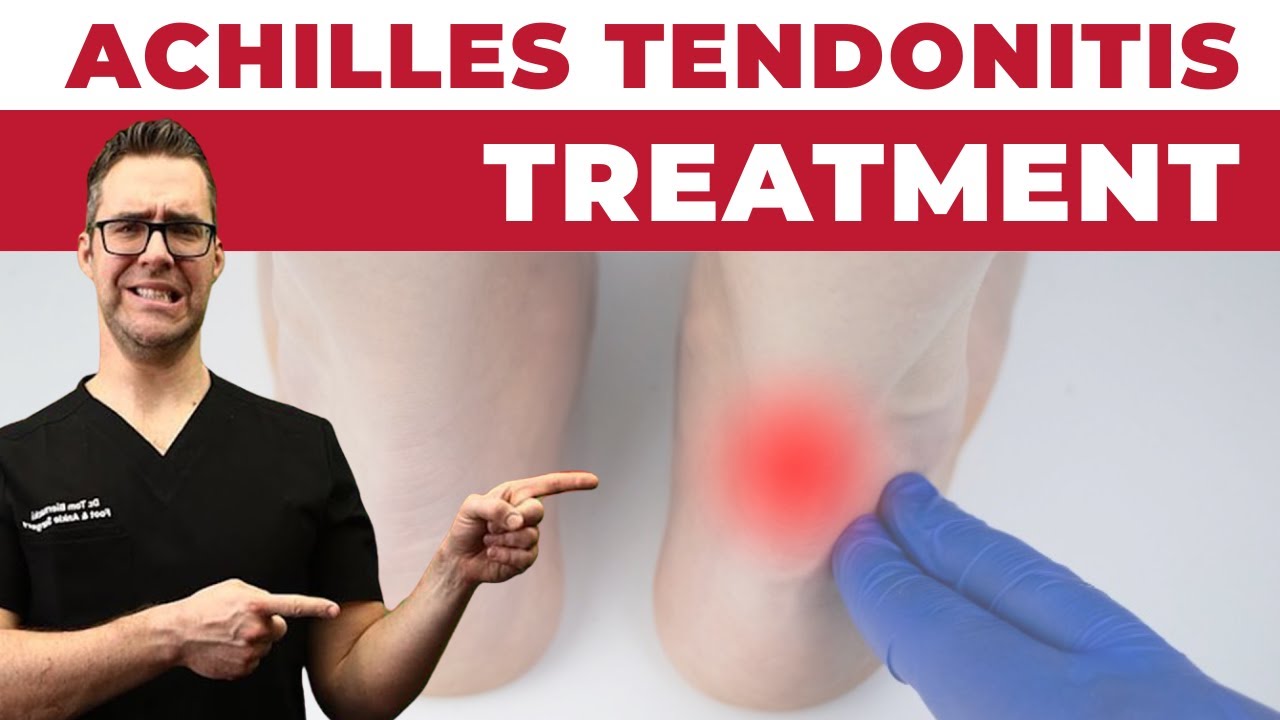
Watch: Achilles Tendonitis Back of Heel Pain — MichiganFootDoctors YouTube
The most important clinical decision with Foot Tendons isn’t which treatment to start with — it’s identifying the correct subtype. That changes everything. Call (810) 206-1402.
Foot Tendons: Quick Answer
The foot has 13 named tendons that cross the ankle joint, divided into 4 functional groups: anterior (extensor) tendons that lift the foot and toes (tibialis anterior, EHL, EDL, peroneus tertius); lateral (peroneal) tendons that evert the foot (peroneus longus, peroneus brevis); deep posterior tendons that invert the foot and flex the toes (tibialis posterior, FDL, FHL); and the superficial posterior Achilles tendon group (gastroc + soleus combining into the Achilles). Plus 4 intrinsic tendon groups within the foot itself (toe flexors, toe extensors, peroneals continued, lumbricals). Knowing which tendon is causing your pain is critical because each requires different treatment — for example, posterior tibial tendinopathy needs medial-post orthotics while peroneal tendinopathy needs lateral wedging.
The 4 Functional Groups of Foot Tendons
Foot tendons are organized into 4 functional groups based on which compartment of the lower leg they originate from:
Anterior (extensor) group: Tibialis anterior, extensor hallucis longus (EHL), extensor digitorum longus (EDL), peroneus tertius. Function: dorsiflexion (lifting the foot up) and toe extension. Innervated by the deep peroneal nerve.
Lateral (peroneal) group: Peroneus longus, peroneus brevis. Function: eversion of the foot. Both run behind the lateral malleolus and insert on the lateral foot. Innervated by the superficial peroneal nerve.
Deep posterior group: Tibialis posterior (TP), flexor digitorum longus (FDL), flexor hallucis longus (FHL). Function: inversion, plantarflexion, toe flexion. All run behind the medial malleolus through the tarsal tunnel. Innervated by the tibial nerve.
Superficial posterior group: Gastrocnemius and soleus muscles join to form the Achilles tendon, which inserts on the calcaneus. Function: powerful plantarflexion (toe-off during gait). Innervated by the tibial nerve.
Tibialis Anterior — The Most Important Anterior Tendon
Origin: Lateral surface of the tibia. Course: Down the front of the shin, over the front of the ankle, across the dorsum of the foot. Insertion: Medial cuneiform and base of the 1st metatarsal.
Function: Primary dorsiflexor of the foot. Lifts the foot during the swing phase of gait. Decelerates the foot at heel strike (eccentric contraction) — preventing foot slap. Inverts the foot.
Common pathologies: Anterior tibialis tendinopathy (worse with running and uphill walking), tibialis anterior rupture (rare, usually in older patients with diabetes or steroid use, presents as foot drop), shin splints (medial tibial stress syndrome — primarily a periosteal reaction but the tibialis anterior is involved). See our anterior tibialis tendonitis guide.
Extensor Tendons (EHL, EDL) — Lift the Toes
Extensor hallucis longus (EHL): Originates from the middle fibula, inserts on the distal phalanx of the big toe. Lifts the big toe. Tested clinically by asking the patient to extend the big toe against resistance (L5 motor function check).
Extensor digitorum longus (EDL): Originates from the lateral tibial condyle, inserts on toes 2-5 distal phalanges. Lifts toes 2-5.
Common pathologies: Extensor tendinopathy from tight shoe lacing, top-of-foot pain in runners. Treatment: window lacing, supportive shoes, NSAIDs. Read our extensor tendonitis guide.
Peroneal Tendons — Lateral Stabilizers
Peroneus longus: Originates from the proximal fibula, runs behind the lateral malleolus, then crosses under the foot to insert on the medial cuneiform and 1st metatarsal. Function: eversion + plantarflexion + supports the lateral arch.
Peroneus brevis: Originates from the distal fibula, runs behind the lateral malleolus, inserts on the base of the 5th metatarsal. Function: eversion + plantarflexion.
Common pathologies: Peroneal tendinopathy (often after ankle sprains), peroneal tendon subluxation (the tendons slip in front of the lateral malleolus during dorsiflexion), peroneal tendon tear (longitudinal split tear of the peroneus brevis is most common — needs MRI). Treatment: orthotics with lateral wedging, NSAIDs, immobilization for 4-6 weeks if severe; surgical repair for refractory cases.
Posterior Tibial Tendon — The Arch Supporter
Origin: Posterior tibia and fibula. Course: Down the back of the shin, behind the medial malleolus, then forward to insert on the navicular and multiple midfoot bones.
Function: The MOST important tendon for arch support. Inverts the foot. Plantarflexes the foot. Locks the midtarsal joint during push-off, transforming the foot from a flexible shock absorber to a rigid lever.
Common pathologies: Posterior tibial tendon dysfunction (PTTD) is the leading cause of adult acquired flatfoot. Stages 1-2 respond to orthotics; stages 3-4 require surgical reconstruction. See our PTTD guide.
FHL and FDL — The Toe Flexors
Flexor hallucis longus (FHL): Often called “the dancer’s tendon” — flexes the big toe at toe-off and during ballet en pointe positions. Common pathology: FHL tendinopathy (especially in dancers); tenosynovitis at the posterior ankle. Read our FHL tendonitis guide.
Flexor digitorum longus (FDL): Flexes toes 2-5. Less commonly injured than FHL.
Achilles Tendon — The Strongest Tendon in the Body
Formation: The gastrocnemius and soleus muscles converge to form the Achilles tendon, which inserts on the posterior calcaneus.
Function: Primary plantarflexor of the foot. Generates the force for push-off, jumping, and sprinting. Bears 6-8x body weight during running.
Common pathologies: Insertional Achilles tendinopathy (where it joins the calcaneus), midportion tendinopathy (2-6cm above the insertion), retrocalcaneal bursitis, Haglund’s deformity (pump bump), Achilles rupture (sudden pop with weakness — surgical emergency). Read our back-of-heel pain guide.
When You Need a Podiatrist for Tendon Pain
Same-week appointment if: pain persists more than 4 weeks despite rest and shoe modification; visible swelling that doesn’t resolve; weakness or inability to perform single-leg toe rise; sudden pop with weakness (suspect rupture — emergent); recurrent ankle sprains (often signal peroneal tendon injury). At Balance Foot & Ankle we offer on-site ultrasound for real-time tendon evaluation, plus MRI orders for deeper investigation.
Frequently Asked Questions About Foot Tendons
How many tendons are in the foot?
13 named tendons cross the ankle joint, plus several intrinsic tendons within the foot itself (toe flexors, lumbricals, etc.). The 13 main tendons are organized into 4 functional groups: anterior (4), lateral peroneal (2), deep posterior (3), and superficial posterior Achilles (forming from gastroc + soleus + plantaris).
What is the most commonly injured foot tendon?
The posterior tibial tendon (PTTD is leading cause of adult acquired flatfoot) and the Achilles tendon (Achilles tendinopathy and rupture). Peroneal tendinopathy is also common after ankle sprains.
What is the strongest tendon in the foot?
The Achilles tendon — the strongest and largest tendon in the entire body. Bears 6-8x body weight during running.
Which foot tendon supports the arch?
The posterior tibial tendon is the primary arch supporter. The peroneus longus contributes to the lateral arch.
How long do tendon injuries take to heal?
Mild tendinopathy: 4-8 weeks with appropriate care. Chronic tendinopathy: 3-6 months. Partial tear: 8-12 weeks. Complete rupture (Achilles): 6-12 months full recovery.
Can foot tendons heal without surgery?
Most tendon injuries heal with conservative care: rest, NSAIDs, immobilization (boot or brace), eccentric strengthening, custom orthotics. Surgery is reserved for complete ruptures, refractory tendinopathy after 6 months, or significant tear identified on MRI.
What is the difference between tendinitis and tendinopathy?
Tendinitis (acute inflammation) is rare. Tendinopathy (chronic degenerative changes) is the more accurate term for most tendon problems — the tendon shows microtears and disorganized collagen, not active inflammation. This is why eccentric strengthening (not just rest + NSAIDs) is the cornerstone of modern treatment.
Related Resources from Balance Foot & Ankle
- Extensor Tendonitis (Top of Foot)
- Anterior Tibialis Tendonitis
- Posterior Tibial Tendonitis (PTTD)
- Medial Ankle Tendons
- Flexor Hallucis Longus Tendonitis
- Achilles Tendinopathy
- Shin Muscles Anatomy
Still Dealing With Foot Tendons?
Same-week appointments at Balance Foot & Ankle in Howell & Bloomfield Township, MI.
Book Your AppointmentFrequently Asked Questions
When should I see a podiatrist?
If symptoms persist past 2 weeks, affect your normal activity, or are accompanied by red-flag symptoms (warmth, redness, swelling, inability to bear weight).
What does treatment cost?
Most diagnostic visits and conservative treatments are covered by Medicare and major insurers. Out-of-pocket costs vary by your specific plan.
How quickly can I get an appointment?
Most non-urgent cases see us within 5 business days. Urgent cases (sudden pain, possible fracture) typically same or next business day.
In-Office Treatment at Balance Foot & Ankle
If home treatment isn’t providing relief for your foot pain, our podiatry team at Balance Foot & Ankle can help with same-day evaluations and advanced in-office care.
Ready to Get Relief?
Same-day appointments available in Howell & Bloomfield Township, MI
4.9★ | 1,123 Reviews | 3,000+ Surgeries
Or call: (810) 206-1402

Dr. Tom Biernacki, DPM is a board-certified foot & ankle surgeon (ABFAS & ABPM) at Balance Foot & Ankle Specialists in Southeast Michigan. With over a decade of clinical experience, he specializes in heel pain, bunions, diabetic foot care, sports injuries, and minimally invasive surgery. Dr. Biernacki is a member of the APMA and ACFAS, and his patient education content on MichiganFootDoctors.com and YouTube has made him one of the most-followed foot & ankle educators on YouTube.