

The most important clinical decision with Peroneal Tendonitis Treatment isn’t which treatment to start with — it’s identifying the correct subtype. That changes everything. Call (810) 206-1402 or book online.
Peroneal Tendon Disorders: Tendinitis vs. Tendinopathy vs. Tear — What’s the Difference?
The terms “peroneal tendonitis” and “peroneal tendinopathy” are often used interchangeably — but they represent different stages of the same condition, with different treatment implications. And both are distinct from a peroneal tendon tear, which may require surgery. Getting the right diagnosis determines whether you need rest and rehab, or imaging and surgical consultation.
| Condition | Pathology | Clinical Presentation | Imaging | Treatment |
|---|---|---|---|---|
| Peroneal tendinitis (acute) | Acute inflammatory response in the peroneus longus or brevis tendon; often following a single overuse event or ankle sprain; early-stage with intact tendon architecture | Lateral ankle and posterior fibula pain; tenderness along the tendon course behind the fibula and below the lateral malleolus; swelling along the tendon sheath; pain with resisted foot eversion; onset correlates with recent activity increase or ankle injury | Ultrasound: tendon sheath effusion (fluid around tendon), mild tendon thickening; MRI: peritendinous edema; tendon structurally intact | Rest (relative — reduce aggravating activity); ice; NSAIDs (short course); functional ankle brace; physical therapy — eccentric loading, peroneal strengthening, proprioception; orthotics for lateral arch support; most acute cases resolve in 4-8 weeks |
| Peroneal tendinopathy (chronic) | Degenerative changes in the tendon substance (not primarily inflammatory); collagen fiber disorganization; intratendinous neovascularization; no acute inflammatory cells on histology; results from repetitive microtrauma without adequate recovery | Chronic lateral ankle pain >6 weeks; pain with activity that partially resolves at rest; tendon may feel thickened or nodular on palpation; morning stiffness; no resolution despite standard rest; snapping sometimes present if subluxation component | Ultrasound: tendon thickening, loss of fibrillar echotexture, intratendinous hypoechoic areas (neovascularization on Doppler); MRI: intratendinous signal change; longitudinal splits possible | Eccentric loading protocol (12-week program); shockwave therapy; PRP injection for resistant cases; aggressive PT; surgery considered for cases >6 months refractory to conservative treatment |
| Peroneus brevis longitudinal split tear | Longitudinal (horizontal) split in the peroneus brevis tendon substance — the most common peroneal tendon tear; often occurs at the fibular groove where the tendon is compressed; frequently associated with ankle instability (chronic lateral ankle sprain) | Chronic lateral ankle pain; snapping behind the lateral malleolus with ankle motion (HIGHLY specific for this diagnosis); pain directly over the peroneus brevis behind the fibula; often history of recurrent ankle sprains; tenderness at the fibular groove | MRI: C-shaped or boomerang morphology of the peroneus brevis (normal = oval); split visible as intratendinous cleft; peroneus longus may bowstring through the split. Ultrasound: dynamic exam shows the split with ankle motion | Small, stable tears: conservative (brace, PT, activity modification); 50-60% respond to conservative treatment. Larger or symptomatic tears: surgical debridement + primary repair (tubularization); groove deepening if fibular groove is too shallow; superior peroneal retinaculum repair if subluxation present; 85-90% good outcomes |
| Peroneus longus tear / os peroneum fracture | Less common; tear at the cuboid tunnel (most vulnerable point for PL); often associated with os peroneum (accessory ossicle in the PL tendon) that can fracture; cuboid avulsion fracture possible | Lateral plantar foot pain (not just ankle — differentiates from PB tear); pain at the cuboid level; pain with forced plantarflexion + inversion; snapping at the cuboid tunnel; os peroneum tenderness | X-ray: os peroneum present ± fracture or displacement (compare bilateral); MRI: PL tear at cuboid tunnel; discontinuity in tendon substance | Os peroneum fracture: immobilization first; surgical excision if painful fragment; PL complete tear: surgical repair or reconstruction depending on tear extent and functional demands |
Peroneal Tendonitis Treatment Protocol: 8-Week Conservative Rehabilitation
| Phase | Timeline | Goal | Exercises / Interventions | Activity Level |
|---|---|---|---|---|
| Phase 1 — Load reduction | Weeks 1-2 | Reduce acute pain and inflammation; protect tendon from further loading | Relative rest from aggravating activities; functional ankle brace (lace-up or stirrup); ice 15 min 3× daily; ankle ROM exercises (pain-free range only); ankle pumps to reduce swelling; NSAIDs if acute (5-7 days maximum) | Walking in functional brace; no running, jumping, lateral cutting; sports that require ankle eversion force eliminated temporarily |
| Phase 2 — Range of motion + isometric strengthening | Weeks 2-4 | Restore full ankle ROM; begin loading the peroneal tendons isometrically | Ankle circles; single-leg balance training (eyes open, then closed); isometric eversion against wall (no movement, just gentle sustained contraction); towel scrunches for intrinsic strength; gentle calf stretching (gastrocnemius + soleus) | Walking tolerated; stairs with handrail; stationary cycling if pain-free |
| Phase 3 — Eccentric peroneal loading | Weeks 4-6 | Rebuild peroneal tendon strength and tissue quality; eccentric loading is the most evidence-supported treatment for tendinopathy | Theraband eversion (concentric + eccentric); single-leg heel raise with eversion emphasis; lateral band walks; BOSU balance; resisted eversion with weight or band; ice after loading sessions | Walk + light jog if pain ≤3/10; no lateral cutting or court sports yet; begin swimming and cycling normally |
| Phase 4 — Functional + sport-specific | Weeks 6-8+ | Full functional recovery; return to sport; prevent recurrence | Lateral agility ladder; single-leg squat; plyometric progressions; sport-specific cutting; custom orthotic with lateral arch support (reduces PL demand); confirm adequate single-leg eversion strength vs. contralateral side | Return to sport with lace-up brace for 3-6 months; return to full activity when pain-free with cutting and plyometrics; brace mandatory for higher-risk activities during return |
Peroneal tendonitis on the outside of the ankle responds to bracing, eccentric exercises, and a slight medial wedge — but the wrong combination can let the tendon worsen instead of heal.
You’re in the right place. Dr. Tom Biernacki, DPM, FACFAS — board-certified foot & ankle surgeon with 3,000+ surgeries — explains exactly what peroneal tendonitis treatment means and what works. Call (810) 206-1402 for same-day appointment at Howell or Bloomfield Township.
Quick answer: Treatment for peroneal tendonitis treatment follows a stepwise approach: 1) conservative care first (rest, ice, supportive footwear, OTC anti-inflammatories), 2) physical therapy and targeted exercises, 3) in-office treatments (injections, custom orthotics) if conservative fails at 4-6 weeks, 4) surgery for refractory cases. Most patients resolve at step 1 or 2. Call (810) 206-1402.
Medically reviewed by Dr. Tom Biernacki, DPM
Board-certified podiatric surgeon | Balance Foot & Ankle
Last reviewed: May 2026
⭐ #1 Brace for Peroneal Tendonitis
Peroneal tendonitis develops when the tendons running along the outside of the ankle are overstressed — usually by repetitive inversion or supination. The Active Ankle T2 limits lateral ankle movement without restricting forward motion, directly reducing the mechanical load on the inflamed peroneal tendons during every step.
PowerStep Pinnacle Arch Support Insole
⭐ Corrects the Root Cause of Peroneal Tendonitis
In our clinic, overpronation is the #1 mechanical cause of peroneal tendonitis. As the arch collapses inward, the peroneals contract forcefully to compensate — eventually leading to tendon damage. PowerStep insoles control subtalar joint motion, relieving the eccentric load that drives this condition.
Peroneal Tendonitis Pain Location: Exactly Where It Hurts, and What Else It Could Be
The peroneal tendons start as muscles on the outer lower leg, dive behind the bony bump on the outside of your ankle, and split from there. Peroneus brevis attaches to the base of the fifth metatarsal, the knuckle you can feel halfway along the outer edge of your foot. Peroneus longus keeps going, turns sharply under the cuboid bone, and crosses the sole of the foot to anchor on the inside. Because those two tendons take different paths, where your pain sits tells us which tendon is irritated.
The three classic pain locations
Behind and just below the outer ankle bone. This is the most common spot. Both tendons share a shallow groove here, held in place by a band of tissue called the superior peroneal retinaculum. Pain here is usually an ache that builds during activity, sometimes with a thickened or puffy feel along the tendon.
Along the outer edge of the foot, toward the base of the fifth metatarsal. This is peroneus brevis at its attachment. It is the location most often mistaken for a fifth metatarsal stress fracture, and telling those apart on an exam room table is one of the more common reasons people end up in our office.
Under the outer arch, near the cuboid. This is peroneus longus where it turns under the foot. It gets confused with cuboid syndrome and with plantar fascia problems, even though the treatment is different for each.
What else causes pain in the same area
A lateral ankle sprain hurts in front of and below the outer ankle bone rather than behind it, starts with a specific twisting moment, and usually bruises. A fifth metatarsal stress fracture is pinpoint tender directly on the bone, hurts when you press one finger on it, and gets worse with hopping. Cuboid syndrome sits further forward on the outer midfoot and often follows an inversion sprain. Sural nerve irritation burns, tingles or feels numb rather than aching, and does not change much with resisted movement.
The single most useful self-check is resisted eversion: with your foot relaxed, try to turn the sole of your foot outward while blocking it with your other hand. If that reproduces the pain behind your outer ankle bone, the peroneal tendons are the likely source. If pressing one finger directly on the bone hurts more than resisted movement does, that points at bone rather than tendon, and that distinction is worth an x-ray rather than a guess.
Taping for Peroneal Tendonitis: KT Tape Step by Step
Taping will not heal a peroneal tendon, and any page that tells you otherwise is overselling it. What taping does well is reduce the inversion load that irritates these tendons, which buys you comfortable walking while the actual healing work of load management and strengthening happens.
Kinesiology tape method
Prep. Skin clean and dry, no lotion. Sit with your foot relaxed and the ankle at roughly ninety degrees. Round the corners of each tape strip so they do not peel.
Strip one, along the tendon. Anchor the first two inches with no stretch on the outer lower leg, about a hand width above the outer ankle bone. Run the tape down behind the outer ankle bone at about twenty five to fifty percent stretch, following the tendon path, then lay the final two inches down with no stretch along the outer edge of the foot toward the base of the fifth metatarsal.
Strip two, the support cross. Take a shorter strip, stretch the middle to roughly fifty percent, and lay that stretched middle directly across your most tender point. Lay both ends down with no stretch so it forms an X over the sore spot.
Finish. Rub the tape briskly for a few seconds so body heat activates the adhesive, and wait about half an hour before exercising in it. Take the tape off if you get numbness, pins and needles, spreading redness or itching. Nerves run close to the surface here and tape should never be the reason a foot goes numb.
When a brace beats tape
For most people with genuine peroneal tendonitis, a lace-up ankle brace with side stays does more than tape does, because it mechanically blocks the inward roll that tape only discourages. Tape is a reasonable choice for sport, for people who react to brace pressure, or as a bridge while a brace is on order. In more severe cases we use a walking boot for two to four weeks to genuinely offload the tendon, then step down to a brace as symptoms settle. If your arch is high and this keeps coming back, the underlying problem is often a foot type that overloads the peroneals, and a custom orthotic with lateral posting addresses the cause rather than the flare.
Massage and Self-Release for Peroneal Tendonitis
Massage helps the muscle, not the inflamed tendon. The peroneal muscle belly sits on the outer lower leg between the knee and roughly the lower third of the shin, and that is the area worth working on with a ball or a roller. Slow passes, moderate pressure, a minute or two at a time.
What to avoid matters more than technique here. Do not dig aggressively over the tendon itself behind the outer ankle bone, especially while it is swollen and warm, and do not do deep cross-friction work over an acutely inflamed tendon. If an area is numb or tingling, stop entirely, because that is nerve territory and pressure will make it worse rather than better.
Peroneal Tendonitis Exercises to Avoid
The exercise list above is what to do. This is what quietly keeps people injured:
Stretching the foot inward. Pulling the sole of the foot inward feels like a stretch because it is, but it puts the exact tendon you are trying to calm down on full stretch. This is the single most common self-inflicted setback we see.
Lateral bounding, cutting drills and side shuffles early in recovery, since all of them load the peroneals in the position that hurts. Running on cambered roads, where the road slope tips one ankle outward mile after mile. Barefoot walking on uneven ground before the tendon has settled. High-repetition calf raises in the first couple of weeks. Fast wobble-board work, where a sudden inward roll is exactly the thing you are trying to prevent.
The rule underneath all of these: if a movement reproduces sharp pain behind the outer ankle bone, it is not building tolerance, it is re-injuring the tendon. Discomfort that settles within a few minutes is fine. Pain that lingers into the next morning means you did too much.
If you are in Livingston or Oakland County and this has been going on more than a few weeks, or if you cannot tell whether you are dealing with a tendon or a bone, that is worth an exam and an x-ray rather than more guessing. Call (810) 206-1402 to be seen at our Howell or Bloomfield Hills office.
Related Conditions
In This Article
- What Is Peroneal Tendonitis?
- Peroneal Tendonitis Treatment: The Step-by-Step Protocol
- When Peroneal Tendonitis Needs Medical Treatment
- Warning Signs That Need Urgent Evaluation
- Frequently Asked Questions
- The Bottom Line
- What is Tendonitis?
- Symptoms and warning signs
- Conservative treatment options
- When is surgery considered?
- Recovery timeline and prevention
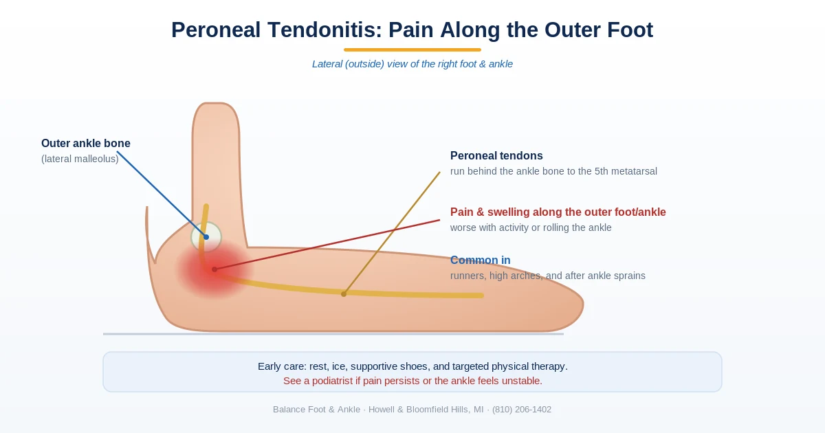
What Is Peroneal Tendonitis?
The peroneal tendons run along the outside of your ankle — the peroneus longus and peroneus brevis. Their job is to evert the foot (roll it outward) and stabilize the ankle during walking, running, and lateral movements. Peroneal tendonitis is inflammation and micro-tearing of these tendons, most commonly at the point where they curve behind the lateral malleolus (the outer ankle bone). In our podiatry clinic, we see peroneal tendonitis most often in runners, basketball and tennis players, and people who’ve recently sprained their ankle. The lateral ankle pain it causes is often mistaken for a lingering ankle sprain — but the treatment is different, and getting it right matters for full recovery.
Peroneal Tendonitis Treatment: The Step-by-Step Protocol
Treatment for peroneal tendonitis follows a predictable progression when managed correctly. The key principle: tendons need controlled loading to heal — complete rest actually delays recovery by preventing collagen remodeling. Here’s the protocol we use in our clinic:
- Phase 1 — Acute (Week 1–2): Relative rest from aggravating activities (no running, cutting, or jumping). Ice 15–20 min 3× daily. NSAIDs if tolerated. Lateral ankle bracing (lace-up or semi-rigid) to reduce peroneal tension. Avoid stretching in this phase.
- Phase 2 — Subacute (Week 2–4): Begin isometric and concentric peroneal strengthening with resistance band — ankle eversion exercises. Cross-training with cycling or swimming. Start lateral wedge orthotic to offload the peroneal insertion.
- Phase 3 — Loading (Week 4–6): Progress to eccentric eversion exercises (the most important phase for tendon remodeling). Single-leg balance work. Begin walking at full intensity if pain-free.
- Phase 4 — Return to Sport (Week 6–8+): Sport-specific agility, cutting, and jumping when eccentric strength is at least 90% of unaffected side. Continue bracing for high-demand activity for 2–3 months after return.
Key takeaway: The most common treatment mistake is complete rest followed by sudden return to activity. Tendons heal through progressive loading — the eccentric strengthening protocol is the evidence-backed core of peroneal tendonitis recovery.
Peroneal Tendonitis Exercises You Can Do at Home
Once the sharpest pain has settled, a simple home program is the core of recovery. The goal is to loosen the tight calf muscles that overload the peroneal tendons and gradually rebuild the strength and balance of the outer ankle. Work in a pain-free range, start with what feels easy, and progress only as comfort allows — mild stretching discomfort is fine, but sharp or worsening pain is your cue to back off.
1. Ankle range-of-motion (start here)
Sitting with the foot lifted, slowly trace the alphabet or draw 10–15 circles in each direction with your toes. This restores gentle motion early without loading the tendon. Do this 2–3 times a day.
2. Calf stretch (gastrocnemius and soleus)
Tight calves are one of the biggest drivers of peroneal tendonitis. Stand facing a wall with one foot back, knee straight and heel down, and lean in until you feel a stretch in the upper calf; hold 30 seconds. Then bend the back knee slightly to shift the stretch lower into the soleus. Three holds each, twice a day.
3. Resisted ankle eversion (the key strengthening move)
The peroneal tendons turn the foot outward, so this is the most important exercise. Sit with a resistance band looped around the outside of your foot and anchored to the opposite side. Keeping the heel still, slowly turn the sole outward against the band, then return with control. Two to three sets of 10–15, building up as it gets easier.
4. Balanced ankle strengthening
With the same band, also work the other directions — pulling the foot up, pointing it down, and turning it gently inward — so the whole ankle strengthens evenly rather than only the injured side. Two sets of 10–15 each.
5. Single-leg balance
Stand on the affected leg for 20–30 seconds, resting a fingertip on a counter for support if needed. As it gets easier, try it with your eyes closed or on a cushion. This retrains the balance reactions of the outer ankle that injury tends to dull. A few rounds daily.
6. Towel scrunches
Sitting with a towel flat on the floor, use your toes to scrunch it toward you. This strengthens the small supporting muscles of the foot that share the load with the peroneals. Two sets.
Work through these most days and expect gradual progress over several weeks rather than days. If the pain isn’t steadily improving after two to three weeks of consistent effort — or if it’s sharp, swelling, or making you limp — that’s the point to have it evaluated rather than push through.
When Peroneal Tendonitis Needs Medical Treatment
Most peroneal tendonitis resolves with conservative care. However, some cases require diagnostic imaging and more aggressive treatment. MRI is the gold standard for evaluating the extent of tendon damage — it can distinguish simple tendonitis from a partial or complete peroneal tendon tear, which changes the treatment approach significantly. Ultrasound-guided cortisone injection can provide relief in chronic cases, though it’s used cautiously near tendons due to rupture risk. Platelet-rich plasma (PRP) injection is an emerging option with promising evidence for chronic peroneal tendinopathy.
Warning Signs That Need Urgent Evaluation
⚠️ See a podiatrist promptly if you have:
- A sudden “snap” or pop on the outer ankle followed by significant swelling (possible tendon rupture)
- A visible bump or lump along the peroneal tendon (tendon subluxation or tear)
- Lateral ankle pain that hasn’t improved after 6 weeks of conservative care
- Ankle that gives way repeatedly with lateral movement (instability)
- Recurrent peroneal tendonitis after multiple episodes (suggests underlying structural issue)
The Bottom Line
Peroneal tendonitis is a highly treatable condition when managed with the right protocol. Relative rest, bracing, and progressive eccentric strengthening resolve most cases in 6–8 weeks. The critical mistake is rushing return to sport before the tendon has adequate tensile strength — this is how acute tendonitis becomes a chronic problem. If you’ve had lateral ankle pain for more than 6 weeks, or if you’ve had multiple episodes of what feels like ankle sprains, a podiatric evaluation including ultrasound or MRI can identify whether you’re dealing with tendonitis, a partial tear, or ankle instability — all of which respond differently to treatment.
Sources: Raikin SM et al. (2024). Peroneal tendon disorders. J Am Acad Orthop Surg. | Van Dijk CN et al. (2023). Conservative management of peroneal tendinopathy. Br J Sports Med.
Dr. Tom’s Picks: Peroneal Tendon Recovery
Apply along the lateral ankle and peroneal groove 3-4x daily. Arnica + menthol reduces tendon inflammation topically. Use during active flares.
View on Amazon →
Graduated 20-30mmHg compression reduces lateral ankle swelling and supports the peroneal tendon sheath. Truly graduated — most ankle braces don’t provide this.
View on Amazon →
Peroneal tendonitis from supination needs arch support to stop the lateral heel strike. Pinnacle repositions the foot and reduces lateral column stress.
View on Amazon →
As an Amazon Associate I earn from qualifying purchases. As a Foundation Wellness partner I may also earn commission.
🦶 Peroneal Tendonitis Recovery Support
- PowerStep Pinnacle Insoles — Peroneal tendonitis is driven by supination (high arches rolling outward) stressing the tendons along the lateral ankle. Correcting that outward roll with the best shoes for supination is a core part of offloading the peroneal tendons. PowerStep corrects the arch mechanics that drive this loading pattern. Medical-grade OTC.
- Doctor Hoy’s Natural Pain Relief Gel — Apply arnica + camphor directly along the peroneal tendons (outer ankle and below lateral malleolus) 3–4× daily. Reduces tendon sheath inflammation.
Peroneal tendonitis that doesn’t respond in 6–8 weeks may have a partial tear. MRI is the diagnostic gold standard. See ankle & tendon treatment options → · Book → · (810) 206-1402
Watch: Peroneal Tendonitis Treatment — What Actually Works

Dr. Tom covers the full peroneal tendonitis treatment protocol — from acute phase bracing and activity modification, through targeted strengthening, to when to consider PRP or surgical reconstruction for peroneal tears. The clinical decision points that separate a 6-week recovery from a 6-month one are clearly explained.
Frequently Asked Questions
How long does peroneal tendonitis take to heal?
Most acute cases settle in 4–8 weeks with relative rest, bracing, and eccentric strengthening. Chronic peroneal tendinopathy — where the tendon has degenerated rather than simply inflamed — can take 3–6 months, and a tendon tear may need imaging and a surgical opinion.
What is the fastest way to heal peroneal tendonitis?
Reduce the aggravating load (not total rest), support the lateral ankle with a brace, and correct the underlying mechanics — usually lateral overload or a high-arched, over-supinated foot — with the right orthotic and footwear. Eccentric peroneal strengthening is the single most evidence-backed exercise.
Should I keep walking with peroneal tendonitis?
Light walking in supportive shoes is usually fine; what sets it back is hills, uneven ground, and worn-out shoes with a broken-down lateral heel. If it hurts along the tendon behind the outer ankle bone while you walk, brace it and book an evaluation.
What shoes are best for peroneal tendonitis?
A stable shoe with a firm heel counter and intact lateral support. Avoid shoes whose outer heel is visibly worn — they tip the foot into the exact position that overloads the peroneal tendons.
What happens if peroneal tendonitis goes untreated?
It can progress from reversible inflammation to chronic tendinopathy, and in some cases to a longitudinal tear or recurrent ankle instability — all harder and slower to fix than the early stage. Persistent outer-ankle pain beyond two weeks warrants an evaluation: (810) 206-1402.
OrthoInfo – AAOS: Peroneal Tendon Injuries
In-Office Treatment at Balance Foot & Ankle
If you’re dealing with peroneal tendonitis pain on the outer ankle and home care isn’t providing lasting relief, our team at Balance Foot & Ankle provides hands-on exam plus imaging when needed and treatment at our Howell and Bloomfield Township locations. We use advanced diagnostic tools to determine the exact cause and build a treatment plan tailored to your activity level and goals.
Same-day appointments available. (810) 206-1402
Peroneal tendonitis that keeps flaring up needs a real diagnosis, not just new shoes — see our ankle & sports injury care. Balance Foot & Ankle’s board-certified podiatrists can help in person at our Bloomfield Township and Howell offices — same-week appointments. Call (810) 206-1402.
Dealing with peroneal tendonitis? See a foot & ankle specialist near you. Balance Foot & Ankle treats peroneal tendonitis and lateral ankle pain — with bracing, physical therapy, custom orthotics, and regenerative options — at our Bloomfield Township office (Oakland County) and Howell office (Livingston County). Book online or call (810) 206-1402.
Clinical sources
- Eccentric Training for Tendinopathies in Athletes: A Scoping Review and Evidence Gap Map. Journal of Sports Science & Medicine. PMC
- Peroneal Tendon Syndromes: Treatment & Management. Medscape / eMedicine. Reference
- Establishing a Conservative Treatment Algorithm for Peroneal Tendon Tears. Podiatry Today. Reference

Dr. Tom Biernacki, DPM is a board-certified foot & ankle surgeon (ABFAS & ABPM) at Balance Foot & Ankle Specialists in Southeast Michigan. With over a decade of clinical experience, he specializes in heel pain, bunions, diabetic foot care, sports injuries, and minimally invasive surgery. Dr. Biernacki is a member of the APMA and ACFAS, and his patient education content on MichiganFootDoctors.com and YouTube has made him one of the most-followed foot & ankle educators on YouTube.